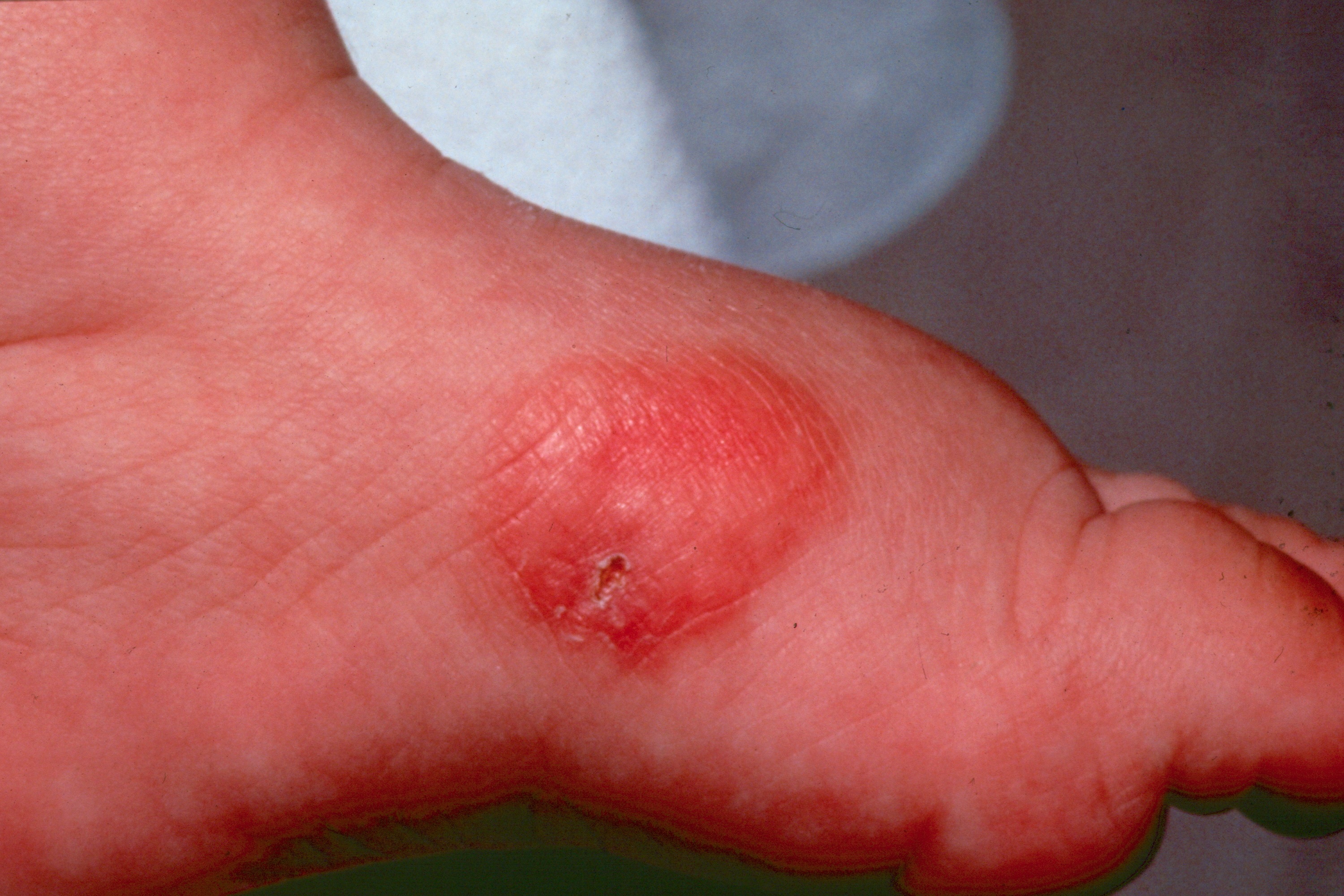
A solitary mastocytoma on the side of the foot of a young child.
SOLITARY MASTOCYTOMA
The solitary mastocytoma (SM) represents an aggregate of dermal mastocytes. It falls within the category of mastocytosis, along with TMEP, urticaria pigmentosa, systemic mastocytosis and bullous mastocytosis.
- Onset is usually within the first 6 months of life.
Clinical
A solitary papulonodule present at birth or the first few weeks of life is most characteristic. It may have an orange peel surface and will urticate (Darier sign) or even blister upon stroking. Rarely, other mastocytomas may form.
Workup
A general physical examination should be done and a CBC obtained.
RegionalDerm
Homepage | Who is Dr. White? | Privacy Policy | FAQs | Use of Images | Contact Dr. White
It is not the intention of RegionalDerm.com to provide specific medical advice, diagnosis or treatment. RegionalDerm.com only intends to provide users with information regarding various medical conditions for educational purposes and will not provide specific medical advice. Information on RegionalDerm.com is not intended as a substitute for seeking medical treatment and you should always seek the advice of a qualified healthcare provider for diagnosis and for answers to your individual questions. Information contained on RegionalDerm.com should never cause you to disregard professional medical advice or delay seeking treatment. If you live in the United States and believe you are having a medical emergency call 911 immediately.