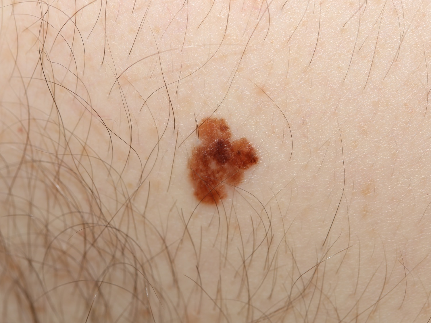
The Melanoma in situ (MIS) is a nevomelanocytic lesion with neoplastic melanocytes limited to the epidermis. Once cells penetrate into the dermis, the lesion becomes a melanoma. The term lentigo maligna (LM) refers to a subset of MIS where a slowly growing pigmented lesion occurs in the sun-exposed areas of an older person. The incidence of MIS has been greatly increasing. For example, in one study, the ratio of MIS:Melanoma was 0.09:1 in 1977 but was 1:1 in 2002.
The usual ABCDE criteria apply here. The MIS most closely resembles the superficial spreading melanoma. Irregular shape, color, dark black areas, etc. are key diagnostic signs. Amelanotic melanoma in situ may occur.
In one study in Connecticut that focused on MIS (either MIS or LM), the MIS to invasive melanoma case ratio during the 5-year period was 257/190 (1.35:1). In that same study, MIS occurred more frequently on the head-neck (119/257) than the trunk (67/257), a reversal of invasive melanoma distribution.
In general, patients should be reassured that although they must be screened yearly, their life expectancy is near normal. The trouble is that rarely, patients will die of "metastasizing MIS". Prompted by the death of a patient with apparent “metastasizing melanoma in situ”, one study reassessed 104 cases of MIS whose diagnosis had been determined with H&E. Immunohistochemical analysis using Mela-A/Mart-1 was employed and showed 30 of 104 (29%) had invasive melanomas. (positive staining cells in the dermis). Most (26/30) had a depth of < 0.5 mm. [The Lancet 2002; 359, 9321;1921]. Another more recent study confirmed this. It found occult invasive melanoma in 33% of previously diagnosed MIS [JAMA Derm 2016;152;1201].
Patients with MIS need annual surveillance for the rest of their life as the risk of developing a second melanoma is elevated and on par with (or in some studies even higher than) patients with melanoma. For example, Youlden et al found that patients with a history of MIS are 4.6 times more likely to develop a subsequent primary melanoma compared to the general population.
Homepage | Who is Dr. White? | Privacy Policy | FAQs | Use of Images | Contact Dr. White