Lentigo maligna. A pigmented patch on the sun-exposed areas of the head, neck, back, or arms of a Caucasian is typical
Lentigo maligna. A pigmented patch on the sun-exposed areas of the head, neck, back, or arms of a Caucasian is typical
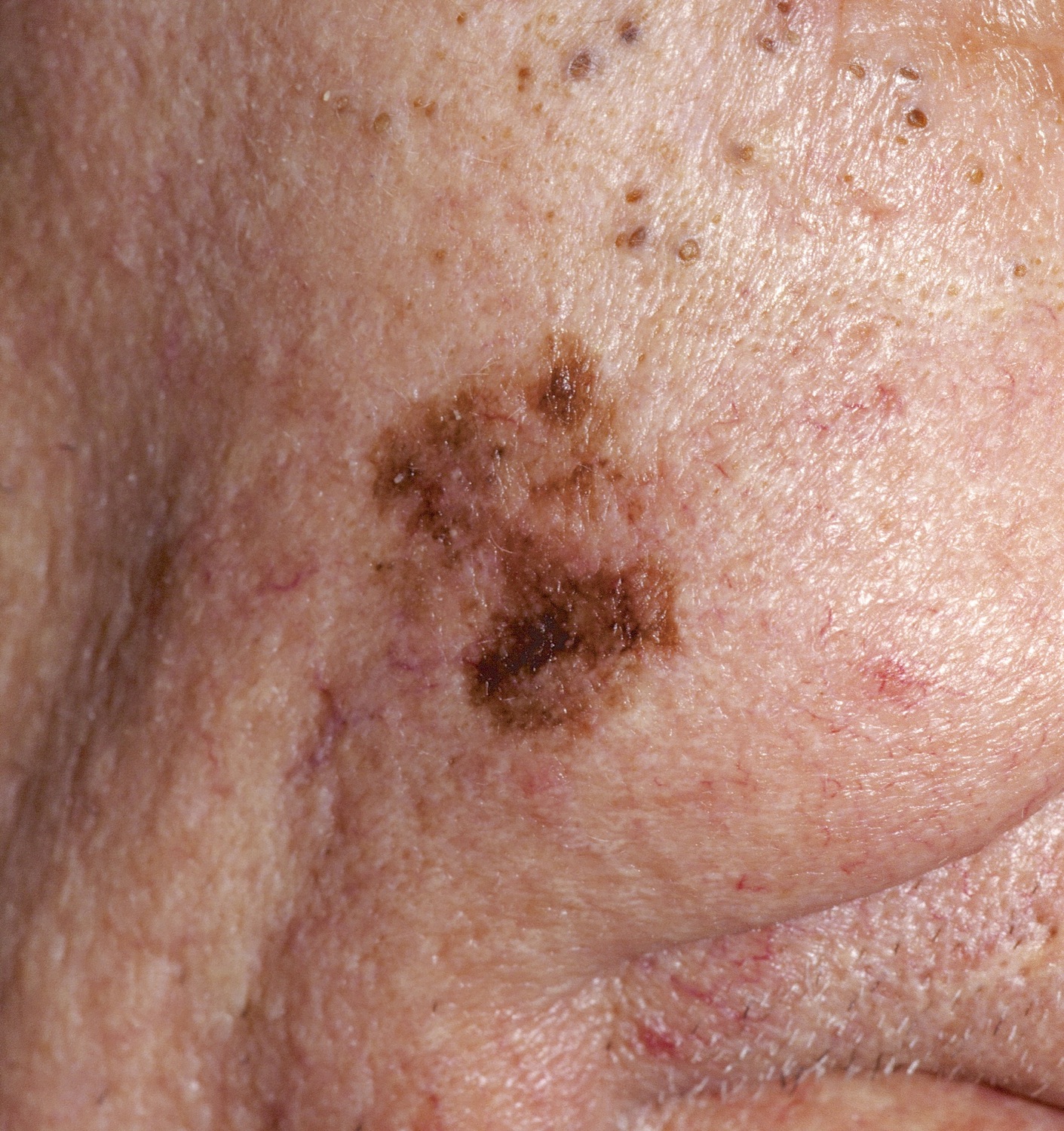
The Lentigo Maligna (LM) is a variant of melanoma in situ that arises in sun-damaged skin in a fair-skinned, older patient. Lentigo maligna may progress to lentigo maligna melanoma (LMM)--true melanoma where invasion has occurred. For LMM, the prognosis is the same as other melanomas based upon depth. It comprises about 5-15% of all melanomas.
A pigmented patch on the sun-exposed areas of the head, neck, back, or arms of a Caucasian is typical. Darkly pigmented areas and irregular shapes are seen. Less commonly, amelanotic lesions may occur, which are red or pink and sometimes scaly. Slow growth over many years is the rule. A SPAK can look very similar.
A recent Dutch study showed the lifetime risk of progression of a LM to LMM (without treatment) is about 2%. Another study showed that the lifetime risk of such progression for a 45 year old is 4.7% and for a 65-year-old, 2.2%.
A broad, thin shave biopsy encompassing all of the lesion is best. At a minimum, a biopsy encompassing all colors of the specimen is necessary. One study demonstrated that about 50% of LM are contiguous with lentigines or pigmented actinic keratoses. Dark color is associated with pigment incontinence, not malignancy. For these reasons, a small partial biopsy (e.g., punch) or just biopsying the darkest area has a high risk of giving a false negative result and should be avoided.
Homepage | Who is Dr. White? | Privacy Policy | FAQs | Use of Images | Contact Dr. White