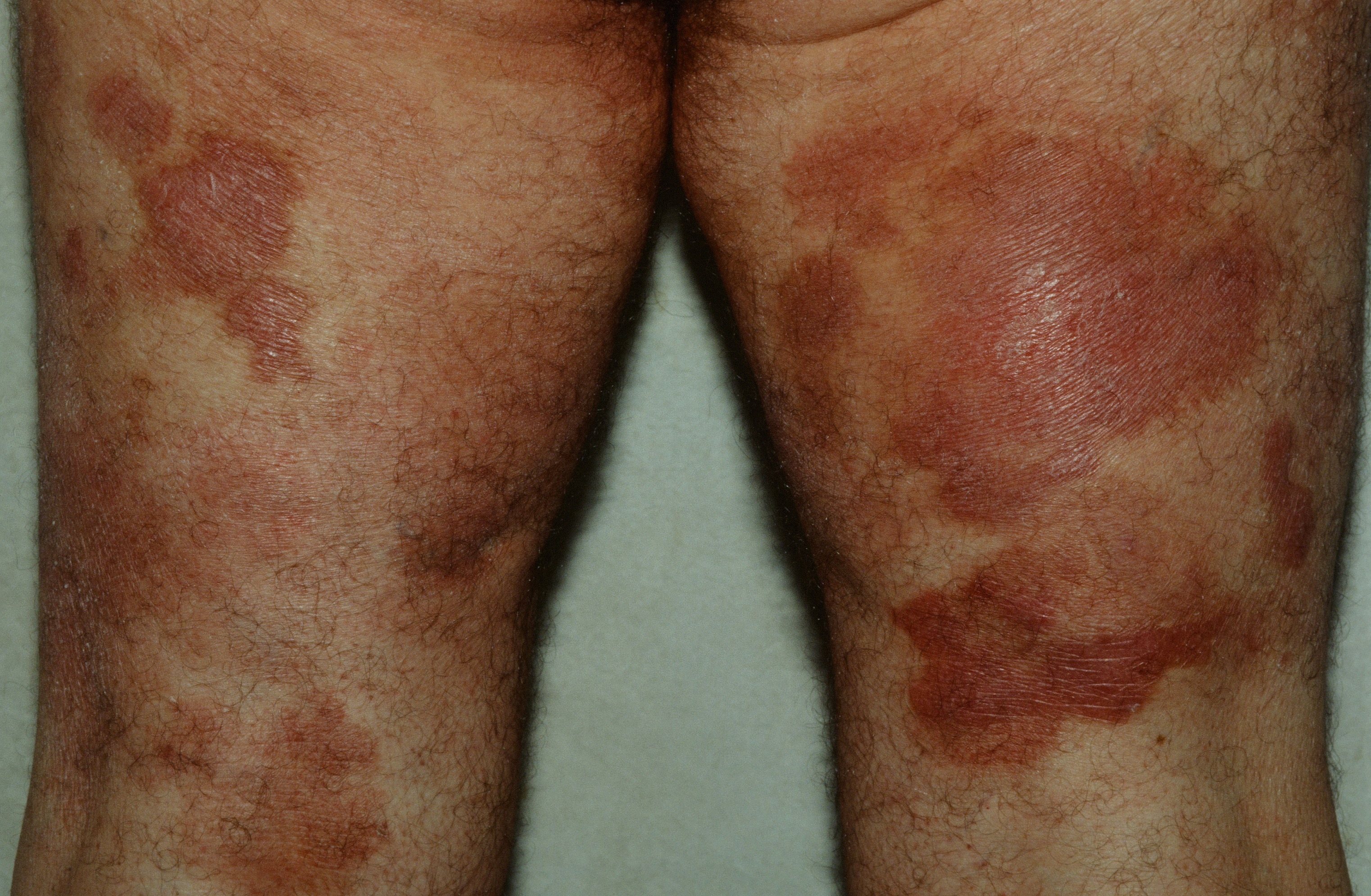
Red/brown, chronic patches and plaques on the upper thighs of an older adult (hidden from the sun). After the appropriate workup, the patient was started on clobetasol, tazarotene and UVB.
Red/brown, chronic patches and plaques on the upper thighs of an older adult (hidden from the sun). After the appropriate workup, the patient was started on clobetasol, tazarotene and UVB.
Mycosis fungoides (MF) and Sézary syndrome (SS) taken together are referred to as Cutaneous T-Cell Lymphoma (CTCL). They represent about 50% of all cutaneous lymphomas. SS is an aggressive, leukemic variant, whereas MF is more indolent.
The patient develops red, scaly areas usually in sun-protected parts of the body, e.g., the thighs, buttocks and legs. Later in the course of the disease, infiltrated plaques, figurate lesions, and nodules may form indicating progression. The development of nodules is a clear sign of more advanced disease. The lesions are fixed in MF in contrast to eczema. Ulceration may occur.
Variants include:
Rarely, children may develop MF. Yellow lesions may indicate xanthomatosis. Vesicobullous lesions may rarely occur. Atypical cases should be evaluated for the presence of HTLV-1 and EBV. Prognosis is unfavorable if tumors, lymphadenopathy, or skin involvement by infiltrated plaques greater than 10% body surface area (BSA) are present. Darker-skinned patients may develop leukoderma from CTCL. Serpiginous CTCL has been reported. Rarely, an elderly patient with diffuse itching but normal skin will show CTCL on biopsy--so called invisible CTCL.
SS has high levels of circulating Sézary cells where as erythrodermic MF (eMF) has low or absent levels. SS arises de novo whereas eMF usually represents progression of MF.
If there is clinical suspicion of CTCL, a skin biopsy should be done which may be diagnostic. If it is equivocal, lymphocyte immunophenotyping may be done. If this is equivocal, gene rearrangement studies may be helpful. Certain drugs can rarely cause a drug eruption mimicking CTCL, so called pseudo-CTCL. A solitary lesion may represent Woringer-Kolopp disease or a response to an arthropod bite.
Workup is usually done in conjunction with an oncologist. For the dermatologist, complete skin exam, calculation of body surface area, and evaluation of lymph nodes should be performed. A biopsy should be taken of any and all morphologies--and certainly of the thickest lesion. The surgical oncologist may want to biopsy any node > 1.5 cm. CXR for limited disease and/or CT scan for widespread/more advanced disease. A drug history should be obtained. On study found hydrochlorothiazide (HCTZ) to be a trigger of CTCL in a small subset of patients. Trial off HCTZ would be in order.
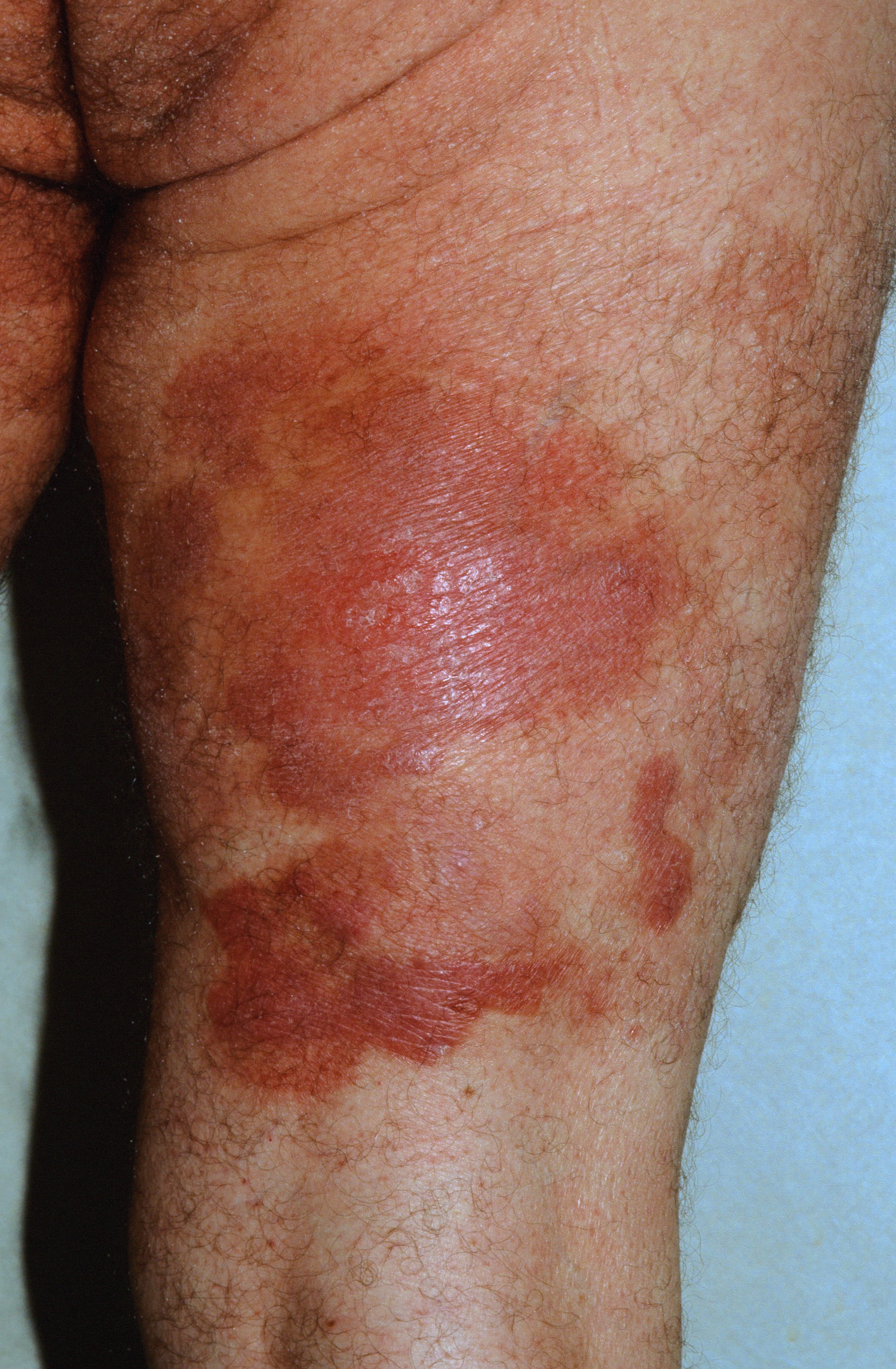
CTCL on the upper thigh.
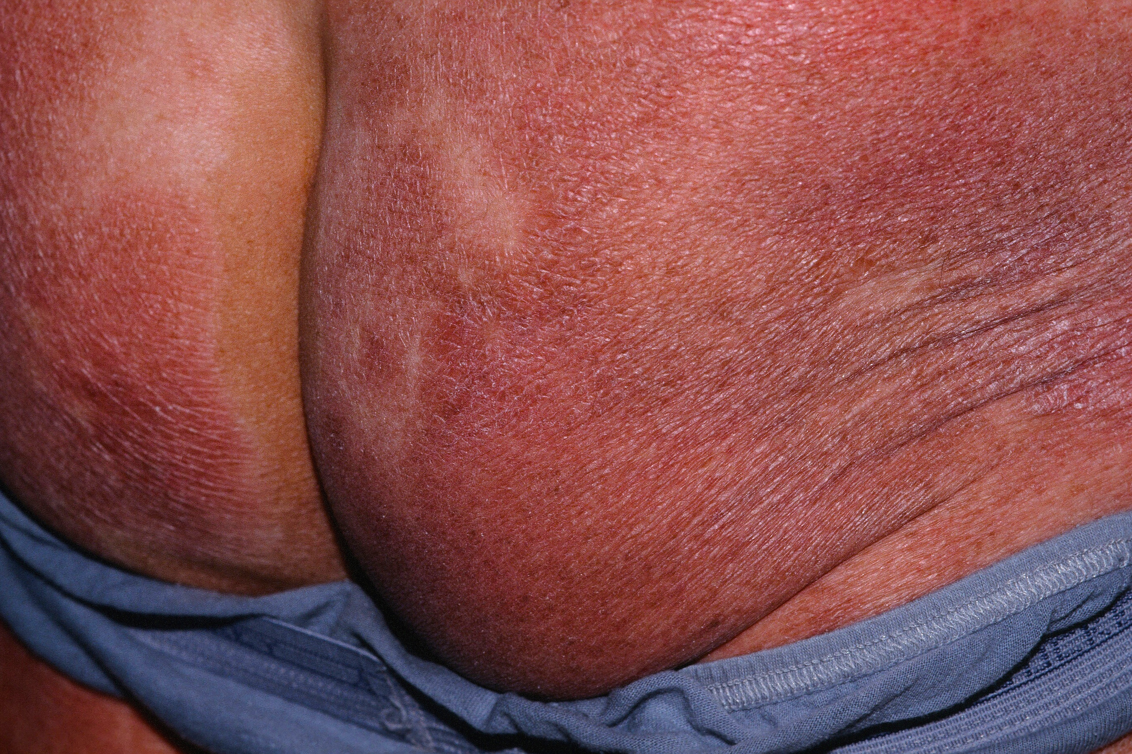
CTCL diffusely of the buttocks.
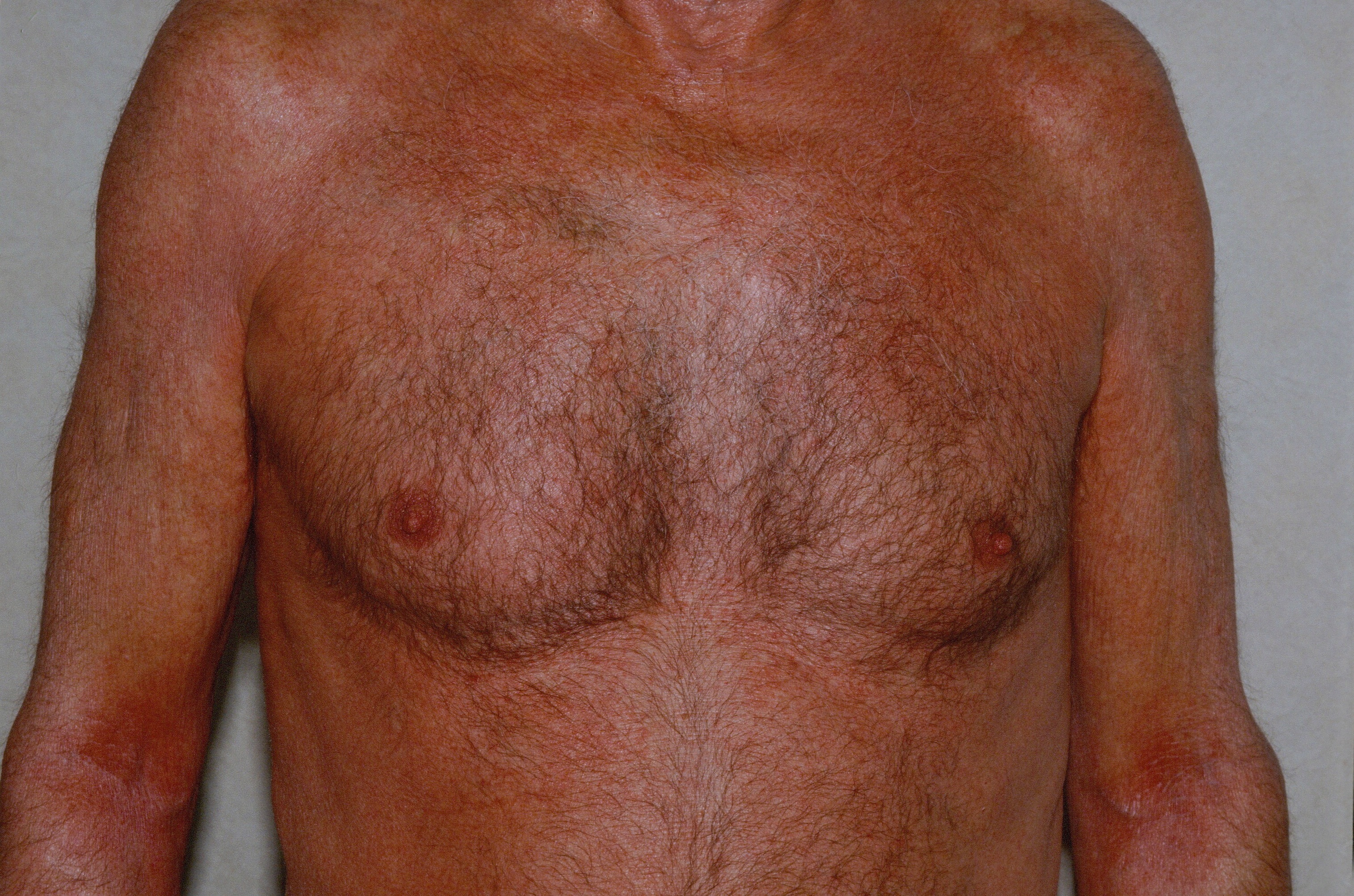
Erytyrodermic CTCL.
Homepage | Who is Dr. White? | Privacy Policy | FAQs | Use of Images | Contact Dr. White