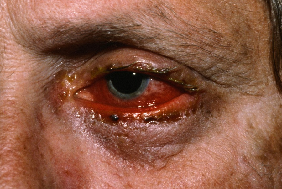
Stevens-Johnson Syndrome (SSS) is a serious allergic reaction in adults characterized by ocular and oral inflammation and diffuse skin reaction or erythema and bulla formation. In children, the term DEN (Drug-Induced Epidermal Necrolysis) is used.
A continuum exists between SJS and TEN with the amount of bulla formation and denudation differentiating the two. For a similar condition in children caused my Mycoplasma pneumonia, see RIME.
The risk of developing SJS varies for individual drugs. In one study [NEJM 1995;333;1600], the highest risk was with sulfonamide antibiotics which conferred a risk of 4.5 cases per million users per week. Trimethoprim-sulfamethoxazole was the sulfonamide most frequently used by affected patients. Other high risk agents included anticonvulsants, cephalosporins, quinolones, tetracyclines, aminopenicillins, NSAIDs and interestingly, systemic steroids. In one study of anticonvulsants [Lancet 1999;353;2190], 16% of cases of SJS or TEN were associated with short term (8 weeks) of use of antiepileptic drugs (e.g. phenytoin, carbamazepine, phenobarbital, and lamotrigine). In that study, valproic acid did not appear to be associated with SJS/TEN.
Inflammation, crusting and redness of the conjunctival, oral and genital mucosa are characteristic in Stevens Johnson syndrome. Headache, fever, malaise and erythema multiforme lesions also occur. The disease occurs most commonly in children triggered by a drug. Inability to eat, fluid loss, and infection are significant complications. These patients may go on to develop TEN. Multiple eruptive melanocytic nevi after Stevens-Johnson syndrome have been reported.
A photo induced SJS has been described. Reported cases have been associated with a ciprofloxicin, sulfasalazine, clobazam, hydroxychloroquine, and naproxen.
It has been asserted that SJS and EM are distinct diseases. A histologic study supported this assertion by finding that severe EM showed a lichenoid inflammatory infiltrate with prominent exocytosis and keratinocyte necrosis limited to the basal layer, whereas SJS demonstrated marked necrosis of the epidermis and only a minimal inflammatory infiltrate with much less exocytosis.
Homepage | Who is Dr. White? | Privacy Policy | FAQs | Use of Images | Contact Dr. White