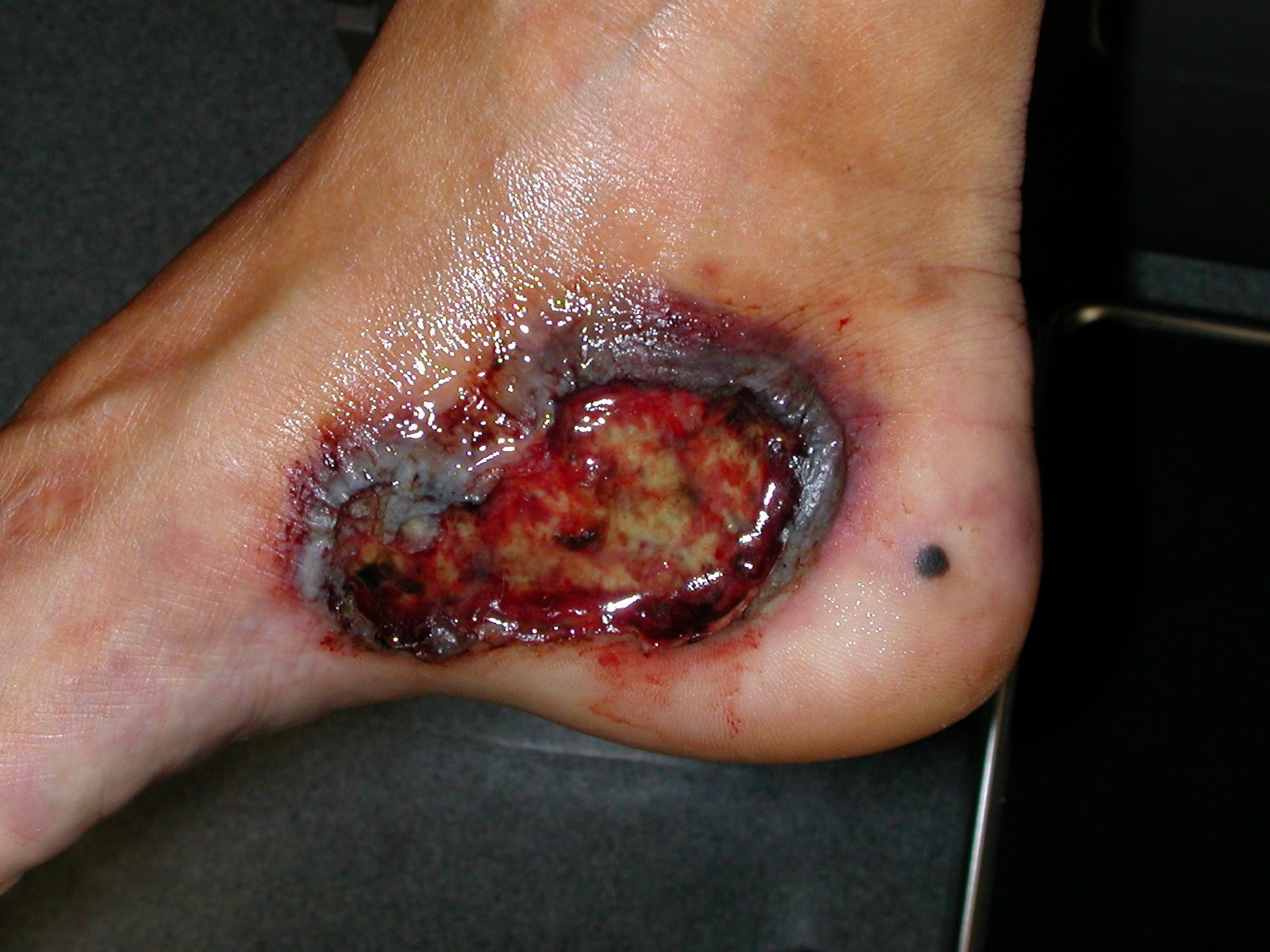
The classic large ulcer with an undermined, violaceous, jagged border.
The classic large ulcer with an undermined, violaceous, jagged border.
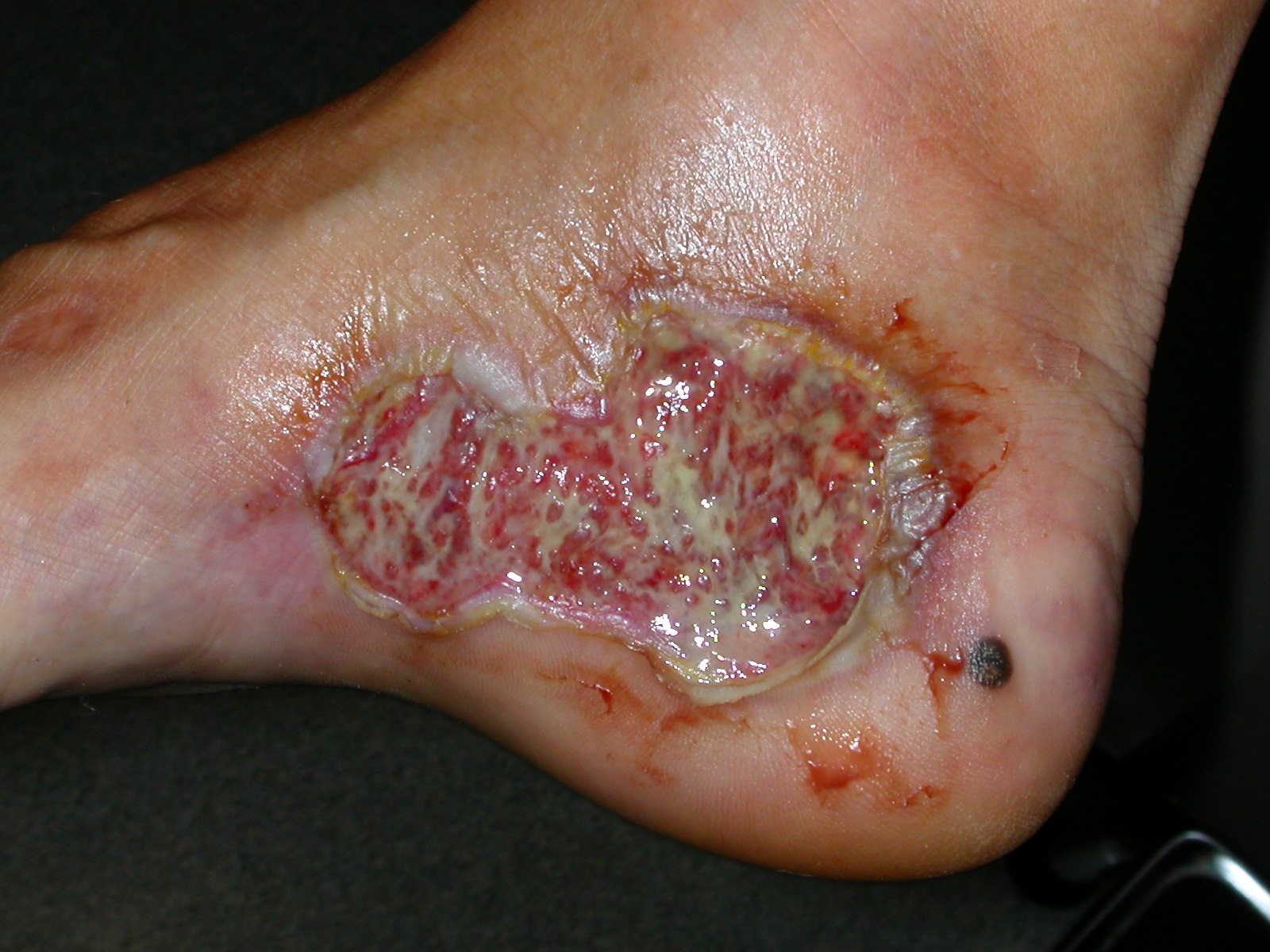
Rapid reduction of inflammation with cyclosporin.
Pyoderma Gangrenosum (PG) is a sterile idiopathic, ulcerative disease that falls within the neutrophilic dermatoses which inclue Sweet syndrome, neutrophilic eccrine hidradenitis Behcet disease, bowel-associated dermatosis-arthritis syndrome, among others. It classically is associated with chronic inflammatory conditions such as ulcerative colitis, Crohn's disease, arthritis, chronic active hepatitis, and Takayasu's arteritis. An IgA paraprotein has been associated in approximately 10% of patients. PG affects slightly more women than men and is most common in patients 10-50 years of age.
PG may be separated into several clinical variants. The classic ulcerative type begins as a papule or pustule which breaks down to form an ulcer with an undermined, violaceous, jagged border. In pustular PG, painful, sterile pustules with a surrounding halo of erythema occur. They may or may not ulcerate. Fever, arthralgias, and the finding of IBD are typical in pustular PG. In bullous PG, pseudovesicular lesions occur. In superficial granulomatous pyoderma, the lesions are more superficial, less aggressive, and more chronic. The edges are less violaceous. There is a predilection for the trunk and many patients do not have any associated diseases. PG lesions of any sort may occur at sites of trauma (pathergy).
An ulcer or ulcers about an ostomy site may represent pyoderma gangrenosum. If Crohn's disease is the reason for the ostomy, involvement of the GIT adjacent to the ostomy site should be considered. Often, there are multiple holes and fistulous tracts that characterize PG as opposed to a periostomal ulcer caused by a poorly fitting ostomy apparatus.
PG has been associated with genodermatoses that involve over activation of the inflammasome, such as PAPA (pyogenic arthritis, pyoderma gangrenosum, acne), PASH syndrome (pyoderma gangrenosum, acne, suppurative hidradenitis), and PAPASH syndrome (pyogenic arthritis, pyoderma gangrenosum, acne, and suppurative hidradenitis)
A review of 13 children with PG found that the skin lesions have a more varied anatomic distribution (i.e., legs, trunk, arms, head and neck), a greater predominance of pustular lesions and a strong association with inflammatory bowel disease--Crohn's disease being the most common.
Drug-induced PG is rare but does occur. Most are colony-stimulatings factor (e.g., granulocyte colony-stimulating factor) or small-molecule tyrosine kinase inhibitors (e.g., gefitinib, imatinib, simitinib and pazopanib).
Extra-cutaneous PG (lung, intestine, cornea, spleen, etc.) involvement occurs. The lungs are the most common site of extracutaneous involvement in PG. A recent study identified pulmonary involvement in 42.7% of cases with systemic involvement. Fever, chest pain, and cough are typically present with pulmonary involvement. Chest imaging studies can show cavitary and/or noncavitary lesions. Ocular involvement was seen in 35.4% of cases. Extracutaneous involvement may occur before, during, or after the appearance of skin lesions
Pyoderma gangrenosum is a diagnosis of exclusion. Diagnosis remains clinical. Exclusion of the many causes of ulcers is necessary. Skin biopsy with cultures for fungal, afb etc is usually needed. One should consider systemic vasculitis (e.g. granulomatosis with polyangiitis), halogenodermas, gummatous syphilis, factitial disease, mycobacterial infection, atypical mycobacterial infection, deep fungal infections, synergistic gangrene, and antiphospholipid syndrome. Occasionally, the PG will abate and yet the ulcers don't heal with continued immunosuppression. Stopping the immunosuppression and good local wound care can heal ulcers in this situation.
A consensus of international experts produced diagnostic criteria [JAMA Derm 2018;154;461] as outlined below. The presence of the one major criteria and 4 of 8 minor criteria maximized discrimination, yielding sensitivity and specificity of 86% and 90%, respectively.
Major Criteria
Minor criteria:
Workup is focused on excluding other disease as PG is a diagnosis of exclusion. It may include ANA, RF, hepatic and renal enzymes, antiphospholipid syndrome, CBC, RPR, SPEP, GI evaluation, serum iodide and bromide levels, and biopsy of the margin of the ulcer for histopathology and culture (e.g., bacterial, viral, fungal, AFB). The patient should be asked about cocaine use as levamisole-adulterated cocaine can precipitate PG. In recalcitrant cases, rarer associations with syndromes should be considered. This may also influence therapy. For example, in cases of PAPA syndrome, PG responds best to IL-1 blockade. Treatment with B vitamins has been shown to improve PG in cases of methylenetetrahydrofolate reductase mutations, whereas patients with myelodysplastic syndrome may improve with thalidomide treatment.
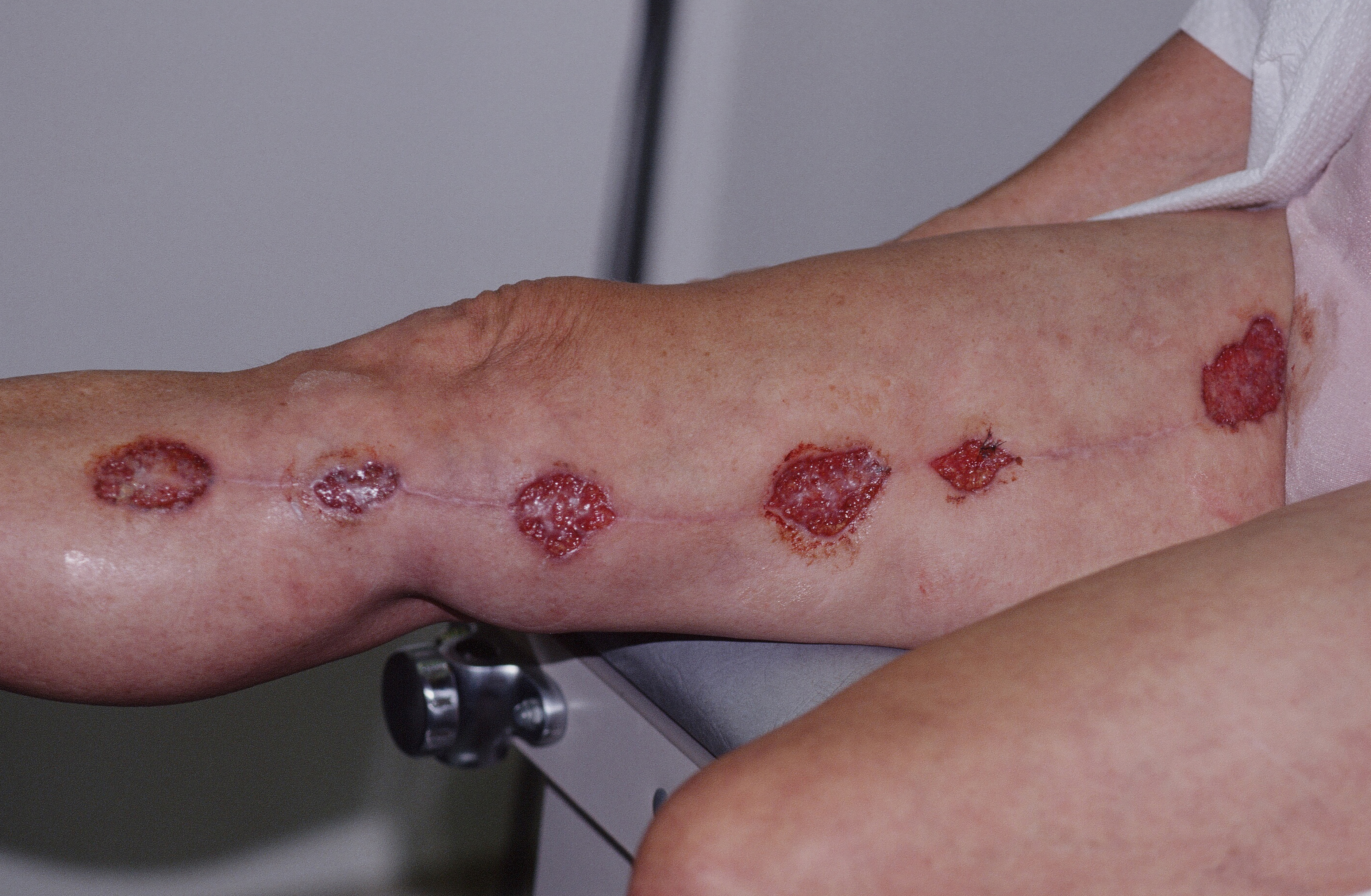
Multiple lesions after surgery.
Homepage | Who is Dr. White? | Privacy Policy | FAQs | Use of Images | Contact Dr. White