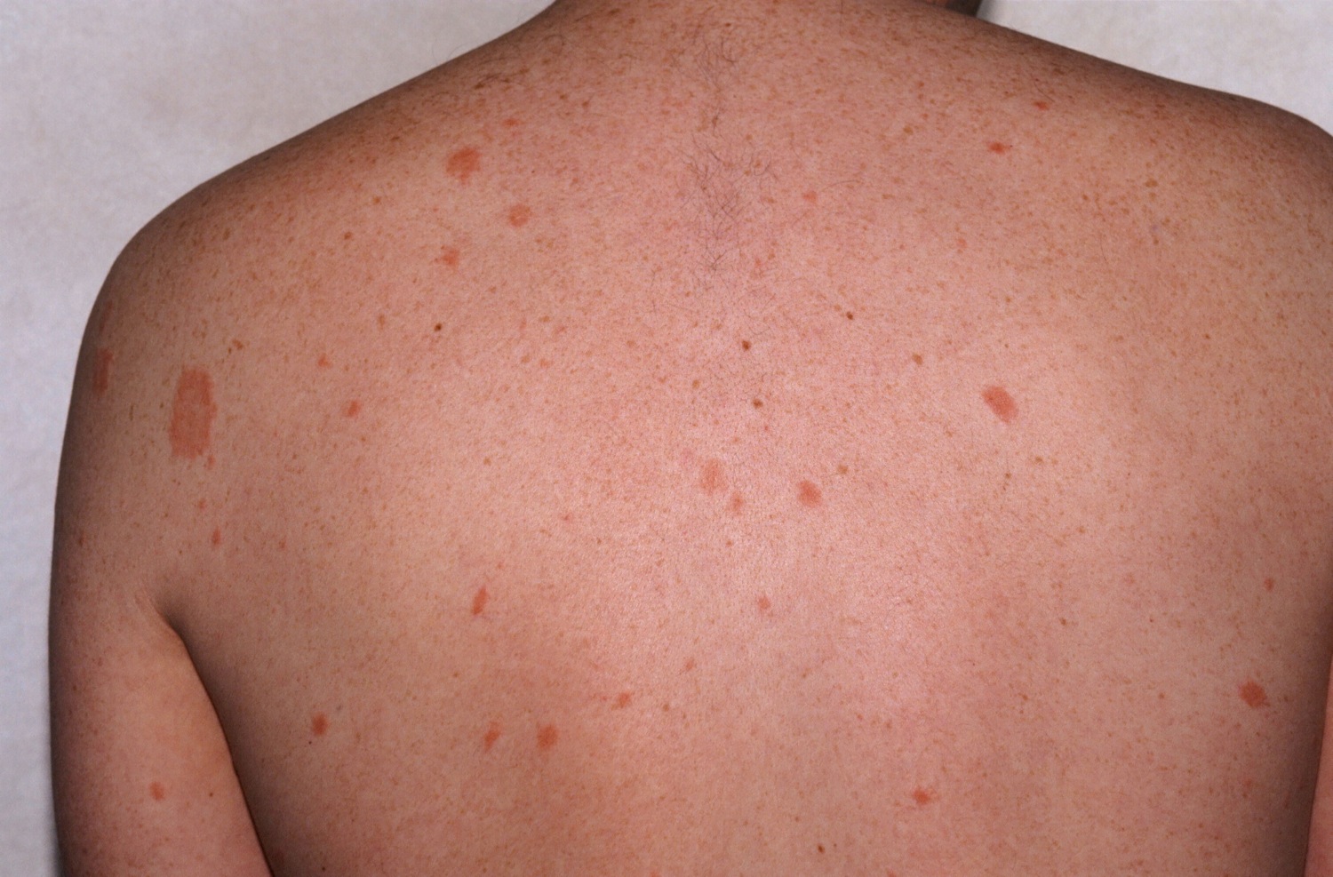
Classic herald patch, the "mother," on the left shoulder, with secondary lesions, the "babies," scattered around on the back.

Classic herald patch, the "mother," on the left shoulder, with secondary lesions, the "babies," scattered around on the back.
Pityriasis rosea (PR) is a common, benign, self-limited papulosquamous skin condition affecting primarily children and young adults. Seventy-five percent of patients are from 10 to 35 years of age.
There is no internal involvement. It is somewhat more common in the winter. Most cases occur in isolation, but occasionally there will be two family members affected. Various drugs have been reported to have caused a PR-like eruption, but it is highly unlikely that a drug can cause the classic presentation of a herald patch followed by secondary lesions.
Probably. Why?
Before the pandemic, there was some speculation that the cause of PR might be a “reactivated” virus and that human herpesviruses 6 and 7 were to blame. Many people become infected at an early age with these two viruses-—both can cause roseola, a childhood skin rash--which then stay in the body. However, PR’s pandemic-related behavior weakened that hypothesis. Cases of PR plummeted during the pandemic suggesting that exposure to some (infectious?) agent is the cause of PR. If reactivation of the roseola virus was the cause, then the pandemic with its assocated stress should have led to more cases.
No increased risk of birth complications and spontaneous abortion was found in pregnant patients with pityriasis rosea compared to matched controls. JAAD 2024
In many patients, a herald patch appears first. The herald patch is round to oval, red and scaly, and may be from 2-6 cm in diameter. It typically occurs on the trunk, neck or proximal extremities. When a patient presents with a solitary papulosquamous lesion of short duration, think PR.
One to several weeks later, the patient experiences a diffuse eruption of red, oval plaques on the trunk, axilla, sides of neck and the groin. PR definitely prefers the sun-protected areas and this is a helpful diagnostic sign. The palms and soles are usually spared (as compared to secondary syphilis where they are usually involved). Many of the plaques may be noted to have a collarette of scale which is also a helpful diagnostic sign. Usually, the long axis of the plaques runs parallel to the ribs giving a Christmas tree pattern.
Oral lesions (exanthema) may occur [JAAD 2017;77;833]. Typical are erythematous macules and papules, vesicles and petechiae especially of the palate.
The areas hidden from the sun like the axilla, buttocks and groin are preferrentially affected. For pictures that are NSFW (not safe for work) see here.
Common considerations are guttate psoriasis, secondary syphilis, drug-induced PR-like eruptions and PLEVA. If there is any question that the patient may have secondary syphilis (e.g., no herald patch and the palms and soles are involved), an RPR should be obtained.
PR and PR-like eruptions have occurred after vaccination including Covid-19. Some eruptions are similar to classic PR with a herald patch and prodromal symptoms. Others are PR-like and do not have prodromal symptoms nor a herald patch. The majority of Covid vaccine PR development derived from the first dose of vaccine. Medium time from vaccine to PR onset was 9 ± 6.3 days. Finally, no cases of severe diseases or PR following the third dose of the vaccines have been described.
PR-like eruptions may occur after drugs including captropril, barbiturates, and isotretinoin.
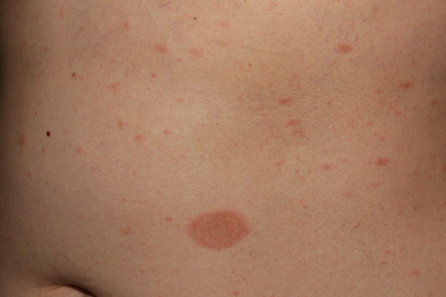
A mother (herald) patch and multiple babies.
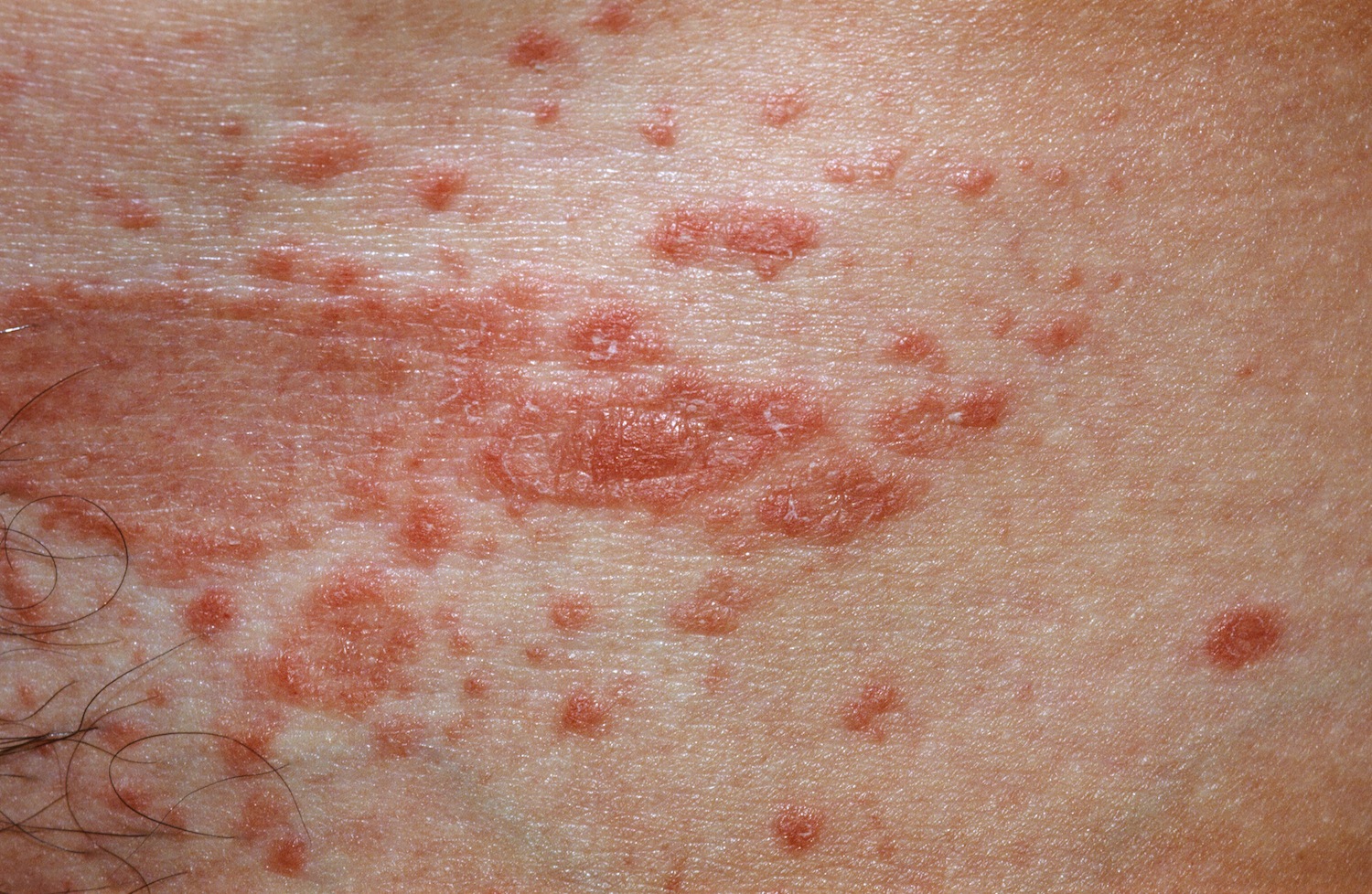
Papules coallescing into plaques in the groin.
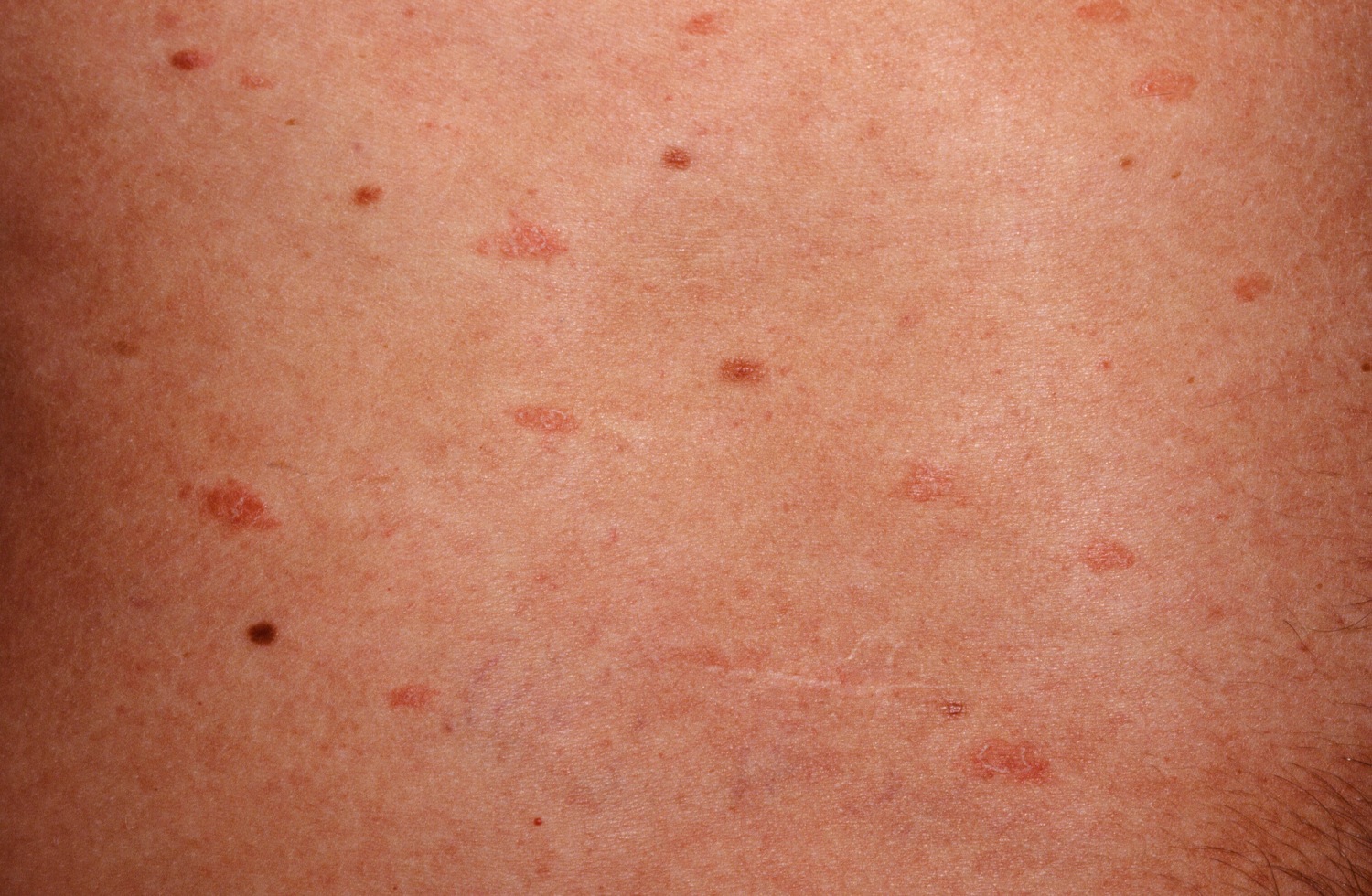
Notice the linear, parallel structure.
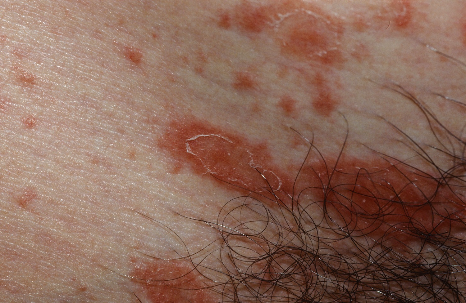 The classic collarette of scale. Notice how the plaque is oriented along the skin lines.
Homepage | Who is Dr. White? | Privacy Policy | FAQs | Use of Images | Contact Dr. White