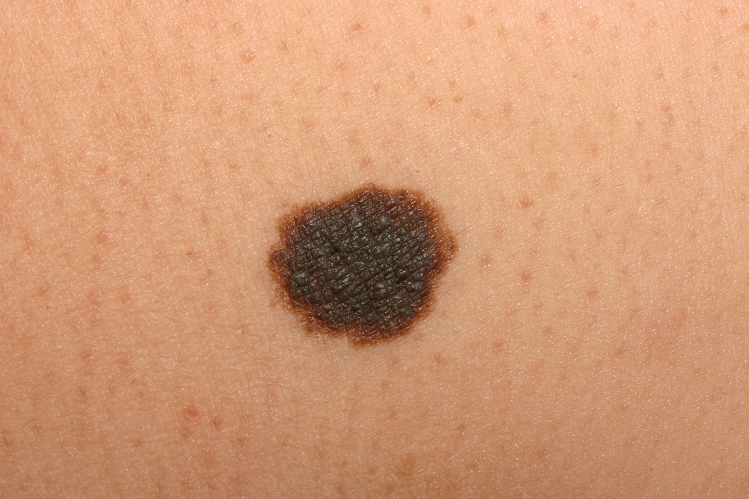
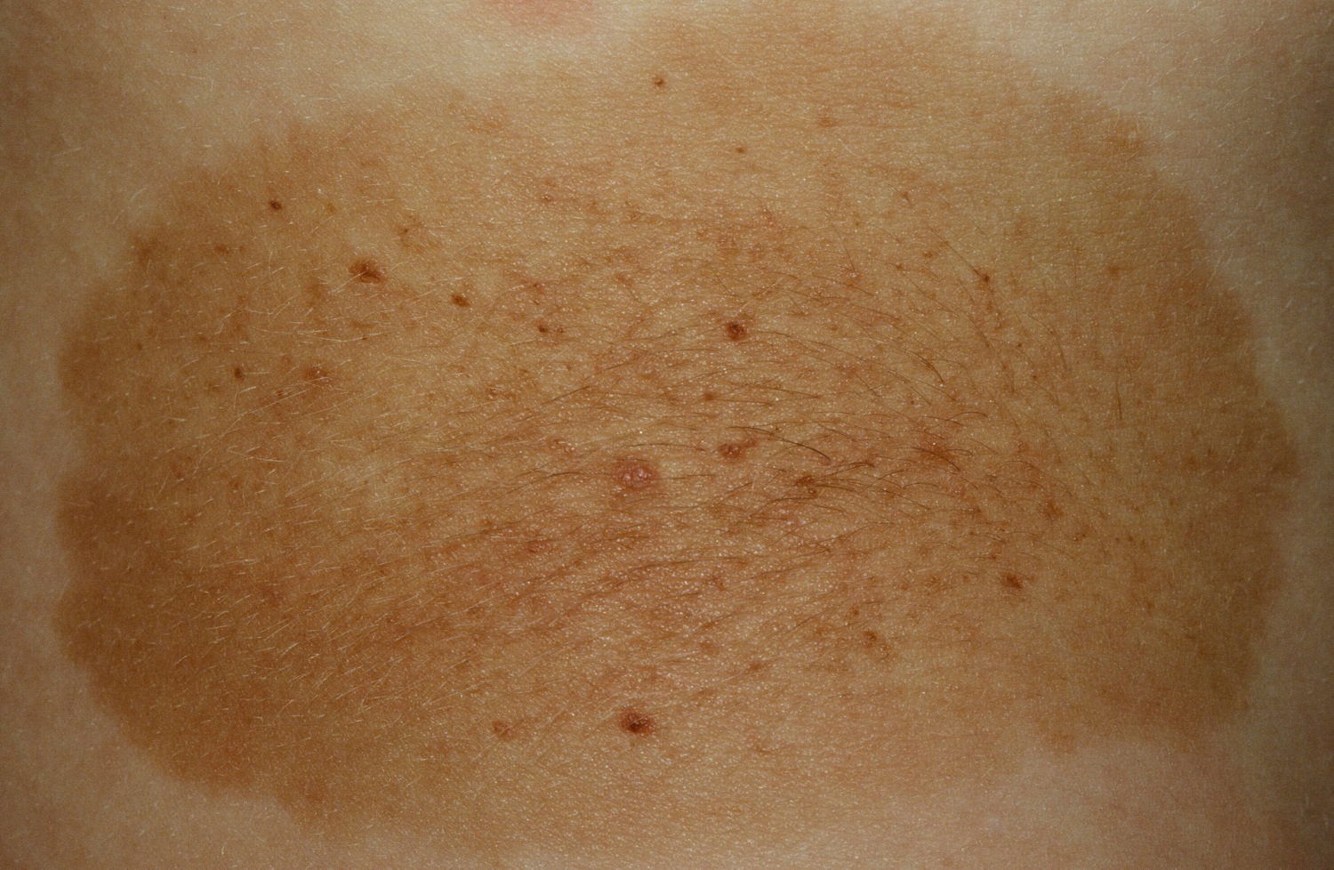
This small congenital nevus has a very low risk for malignant change.
This small congenital nevus has a very low risk for malignant change.
The congenital melanocytic nevus (CMN) is a melanocytic lesion present at or soon after birth.
| Type | Diameter (in an adult) | Frequency | Lifetime Risk of Melanoma |
|---|---|---|---|
| Small | < 1.5 cm | Common | < 1% |
| Medium | 1.5-19.9 cm | Uncommon | < 1% |
| Large | >= 20 cm. | Rare | 5-6%. |
| Giant | > 40 cm | Rare | 5-6% or higher. |
| Location | Diameter |
|---|---|
| Head | 12 cm |
| Hands, feet, torso, forearms, arms, and buttocks | 7 cm |
| Thighs | 5.8 cm |
| Legs | 6 cm |
In the newborn, the congenital nevus is often flat, resembling a cafe au lait macule. Later, it usually thicken slightly and develops a mildly irregular, verrucous surface. Hypertrichosis with terminal hairs occurs in up to 75%. Commonly smaller, darker macules or papules may develop in the center of larger lesions. Sometimes, a pigmented lesion at birth is not diagnosable clinically. Biopsy may show such lesions to be melanocytic nevi, lentigines, cafe au lait macules, or non-specific fibrosis with increased capillaries.
CMN may occur anywhere on the body, even rarely in the mouth. Hypoplasia of the underlying fat may occur. CMN of the scalp may be associated with cutis verticis gyrata.
The risk of development of melanoma in the small and medium sized nevi is small--less than 1%. For example, one study of 227 medium-sized congenital nevi with an average follow up of 6.7 years did not find any developing melanoma.
The lifetime risk of melanoma in patients with LCMN is probably 5% or less.
For large lesions over the scalp, the possibility of associated leptomeningeal melanocytosis should be considered. MRI is helpful in detecting this feature. Large melanocytic nevi over the spine may be associated with a tethered cord. MRI may be used here as well. Two or more congenital melanocytic nevi of any size are also associated with neurocutaneous melanosis and/or melanoma. It has been recommended that any child born with two melanocytic nevi should have an MRI to check for brain involvement within the first 6 months of life.
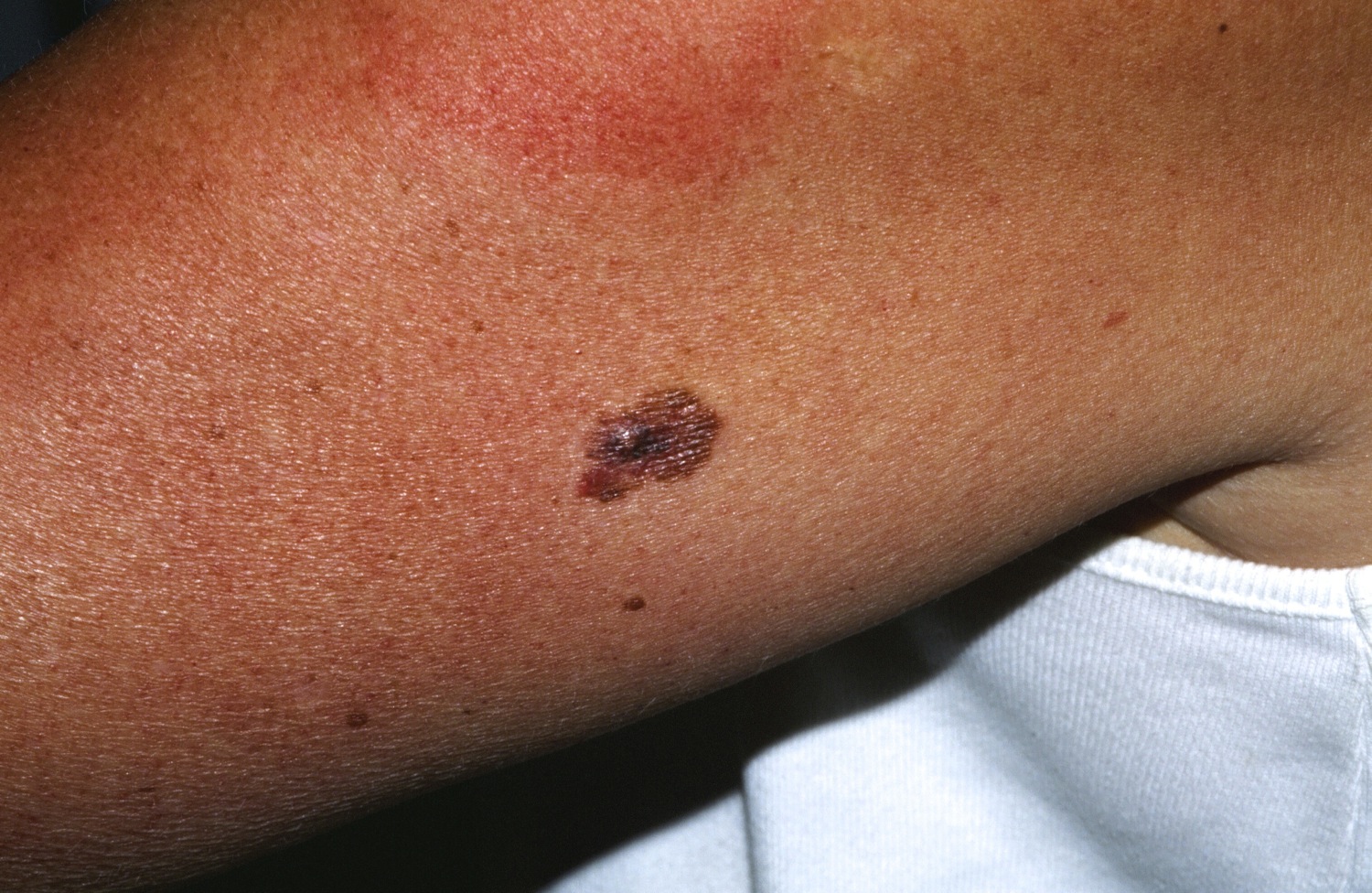
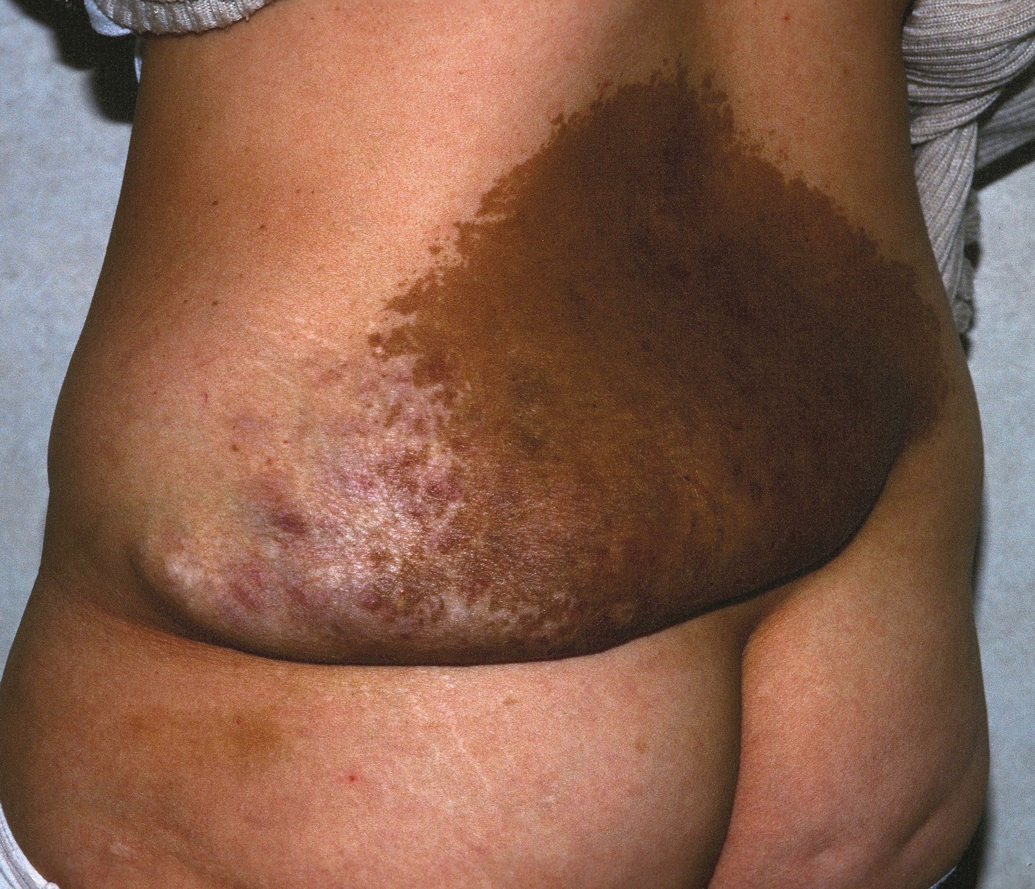
Melanoma developing in a congenital nevus. Although rare, it does happen.
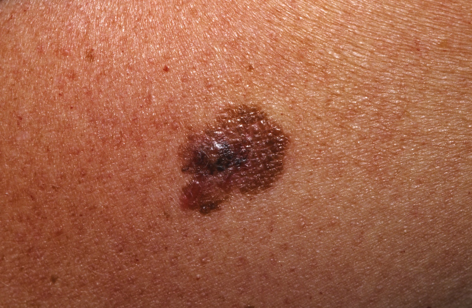
Melanoma developing in a congenital nevus.
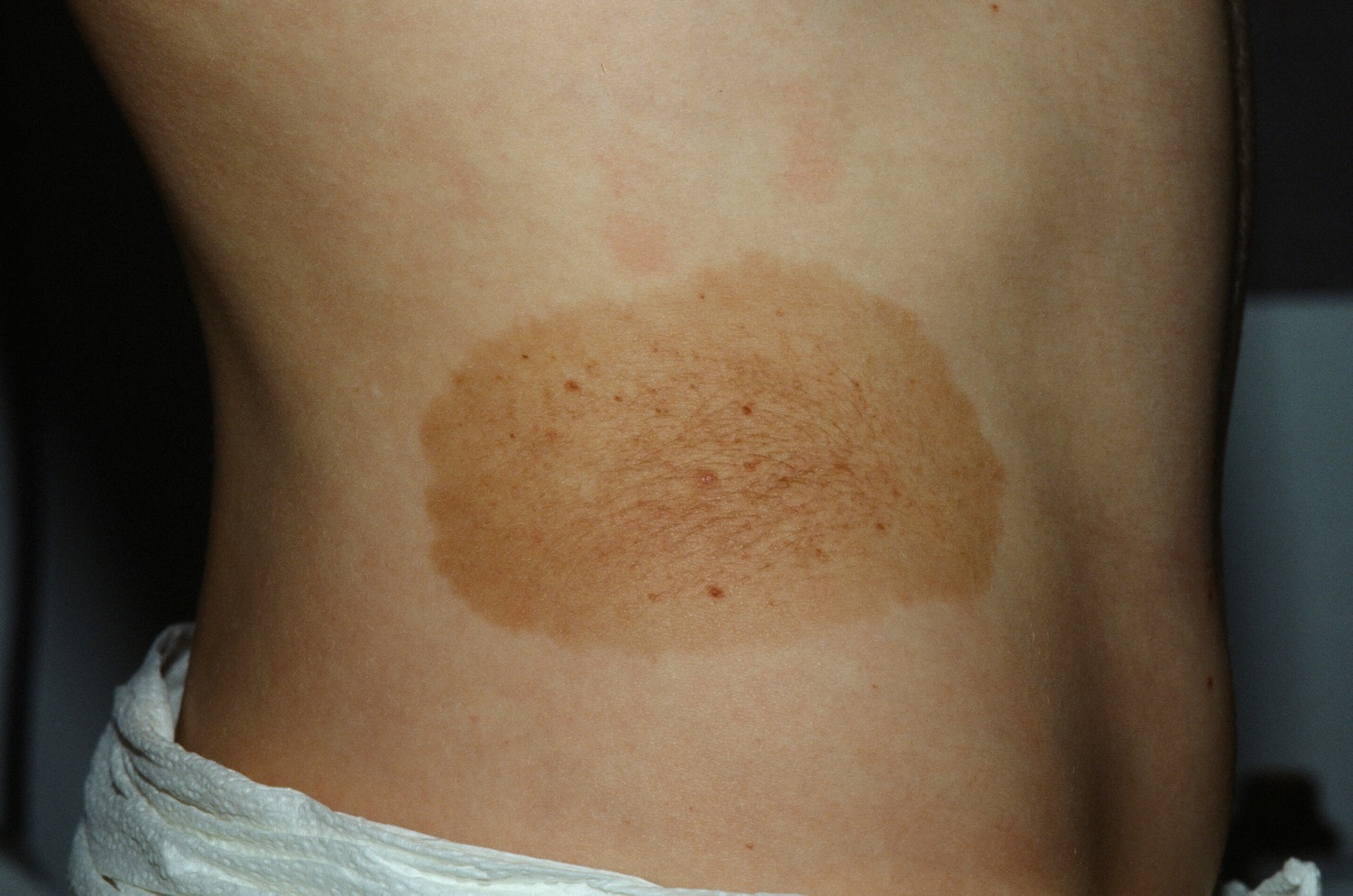
Medium-sized congenital nevus
Congenital Nevus with large Lipoma. Imaging of the spine should be done.
Homepage | Who is Dr. White? | Privacy Policy | FAQs | Use of Images | Contact Dr. White