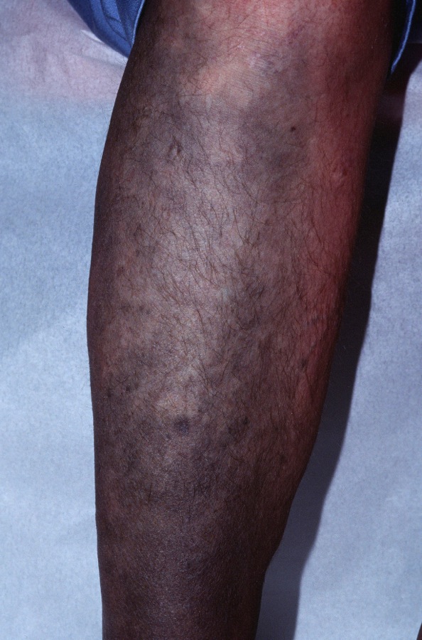
Dark bluish-gray patches on the shins after years of minocyline exposure.

Dark bluish-gray patches on the shins after years of minocyline exposure.
Minocycline Pigmentation (MP) is the development of blue spots or a bluish discoloration of the teeth, nails or sclera in a patient on longterm minocycline. Minocycline is commonly given for acne, rosacea and for rheumatoid arthritis.
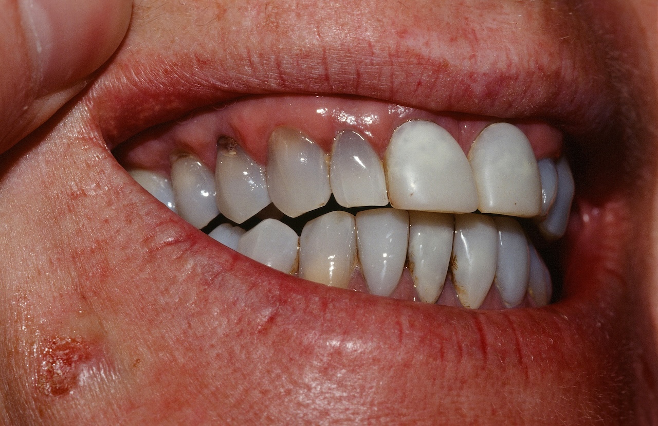
Blue discoloration of the skin may develop on the legs, feet, face, in acne scars, in sclera, and diffusely during minocycline therapy. It may occur as early as the first year of therapy. The teeth, sclera, and nails may also develop a blue-gray discoloration. The gums may appear involved but this actually results from pigmentation of the underlying bone. Pigmentation of bones, thyroid, substantia nigra, atherosclerotic plaques, and conjunctival cysts have been reported. Pefloxacin, a quinolone antibiotic, has caused similar changes on the shins.
Various theories have been proposed about the pigment including: an insoluble minocycline-melanin complex, a complex of minocycline chelated with iron or a drug metabolite-protein complex chelated with calcium. Histopathology shows brown dermal pigmentation that is positive with iron and melanin stains.

Bluish discoloration of the teeth after years of taking minocyline.
Homepage | Who is Dr. White? | Privacy Policy | FAQs | Use of Images | Contact Dr. White