A 30-year-old woman comes to see you complaining of hair loss. This picture shows significant alopecia. Most of the time however, the loss is not so obvious. The patient's history should be trusted.
A 30-year-old woman comes to see you complaining of hair loss.
This picture shows significant alopecia. Most of the time however, the loss is not so obvious. The patient's history should be trusted.
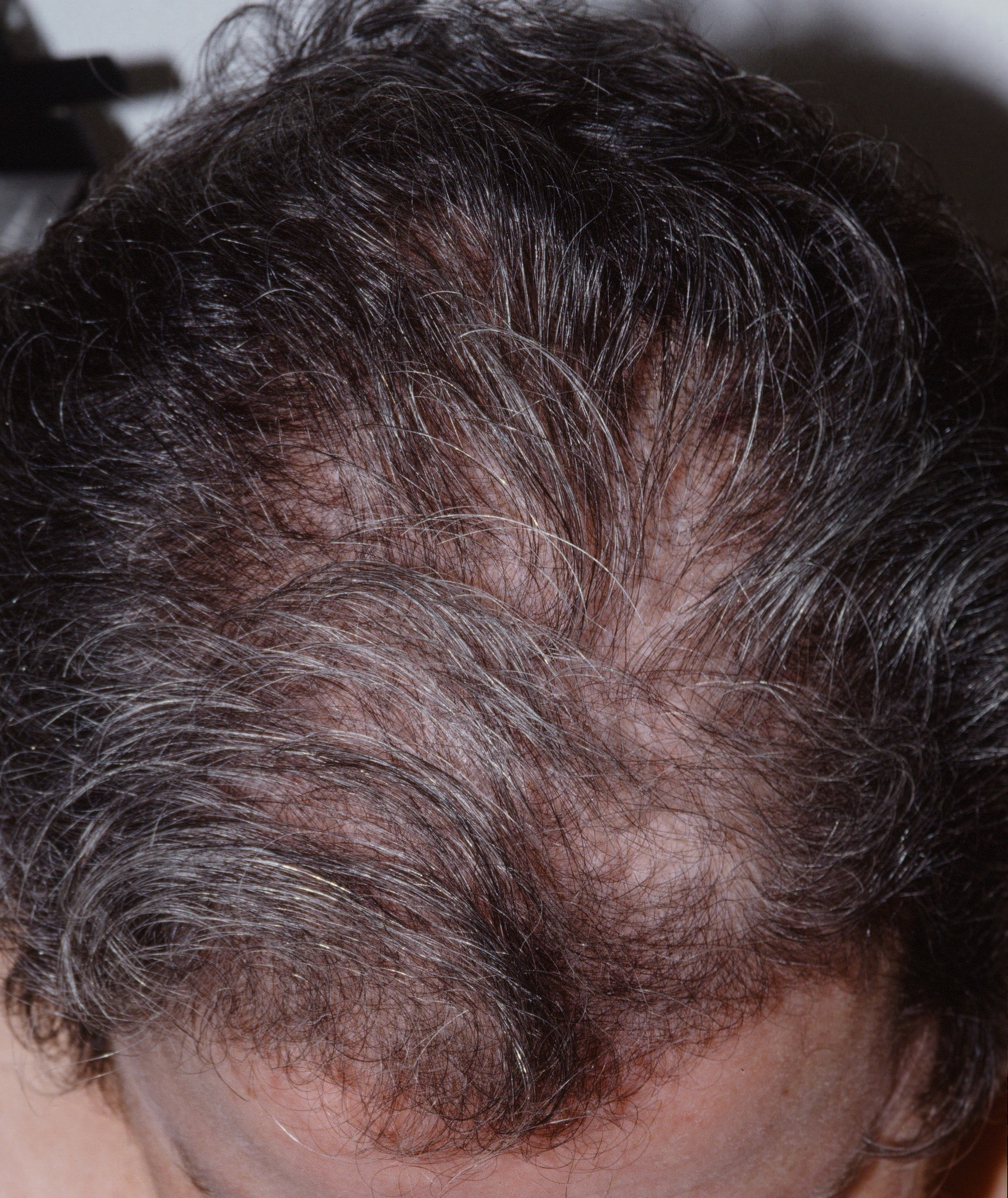
Diffuse hair loss commonly occurs in a young woman. The most common causes are low iron and telogen effluvium.
This page discusses the clinical situation of a young to middle-aged woman with diffuse hair thinning/loss. There are no individual bald spots (e.g., as in alopecia areata), inflammation, nor scarring. It does not have the obvious clinical presentation of Frontal Fibrosing Alopecia, Lichen Planopilaris, or Central Centrifugal Alopecia. The onset may be gradual over weeks to months or even years. The loss is usually across the top, sides, and front. Often, the back of the scalp is relatively spared. The skin of the scalp is normal. Much of the time, you as the clinician cannot appreciate the hair loss. In that case, trust the patient!
Most cases can be appropriately managed with history, clinical examination and laboratory studies. However, some cases may require biopsy for diagnosis. A 4 mm punch is preferred, and go 4 mm deep as well. The punch should be oriented at the same angle as the emerging hairs to avoid transecting hair follicles. More than one punch biopsy may be necessary allowing for both horizontal and transverse sectioning. Biopsying the advancing edge is less likely to be helpful than biopsying a more established area. The area chosen should be of several months duration and still active. If not diagnostic, a central scarred area may be biopsied. Evaluation with elastic tissue stains and polarized microscopy is particularly helpful here.
The typical patient is a young to middle-aged woman who complains of hair thinning. Often, physical examination shows what appears to be a full head of hair, but the woman is certain this is abnormal for her. Usually, this is true. Whether the hair thinning is abnormal or a normal process of aging must be discerned. (Most people experience some thinning of hair as they age.) First, an examination should be done to exclude localized alopecia or scarring alopecia. Also, the hair should be falling out from the root, not breaking (as might occur if the hair has been damaged). Then, one should investigate, by history, if there are any acute stresses on the body that might trigger telogen effluvium in the three months leading up to onset (e.g., flu or other more severe illness, death in the family, pregnancy, car accident). Blood loss via heavy periods or blood donation is a common cause of low iron. Finally, the above labs may help exclude internal causes such as hypothyroidism, low iron, a hormonal abnormality and vitamin D deficiency. If the history and physical examination are unremarkable and the labs are normal, the diagnosis is female pattern hair loss (FPHL). Telogen effluvium (TE) may rarely become chronic and differentiation from FPHL may be difficult. The following may be helpful: The onset of FPHL usually is gradual whereas that of TE is more acute. Many miniaturized hairs are present in alopecia areata.
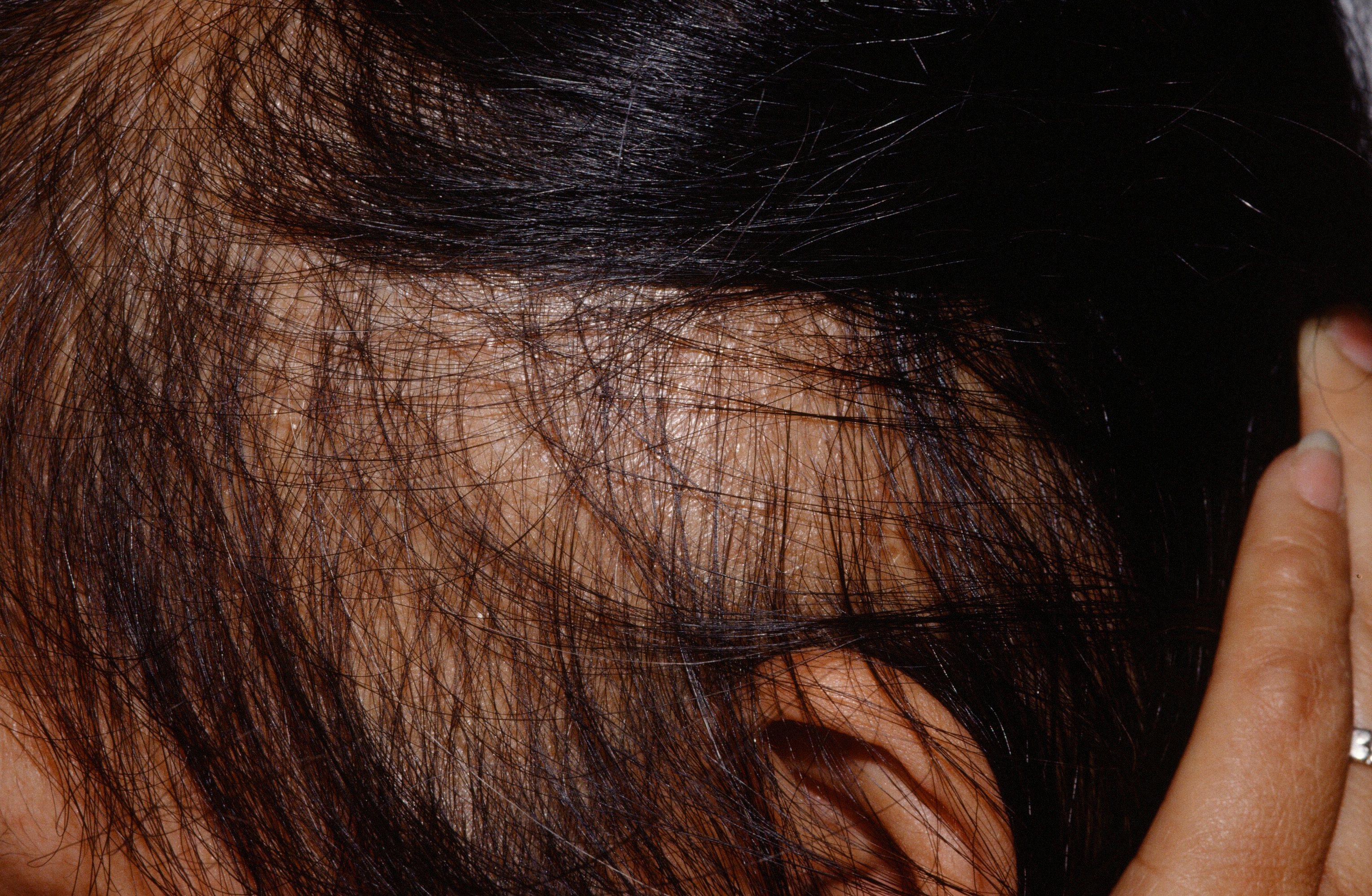
This woman's DHEA was 10 times normal.
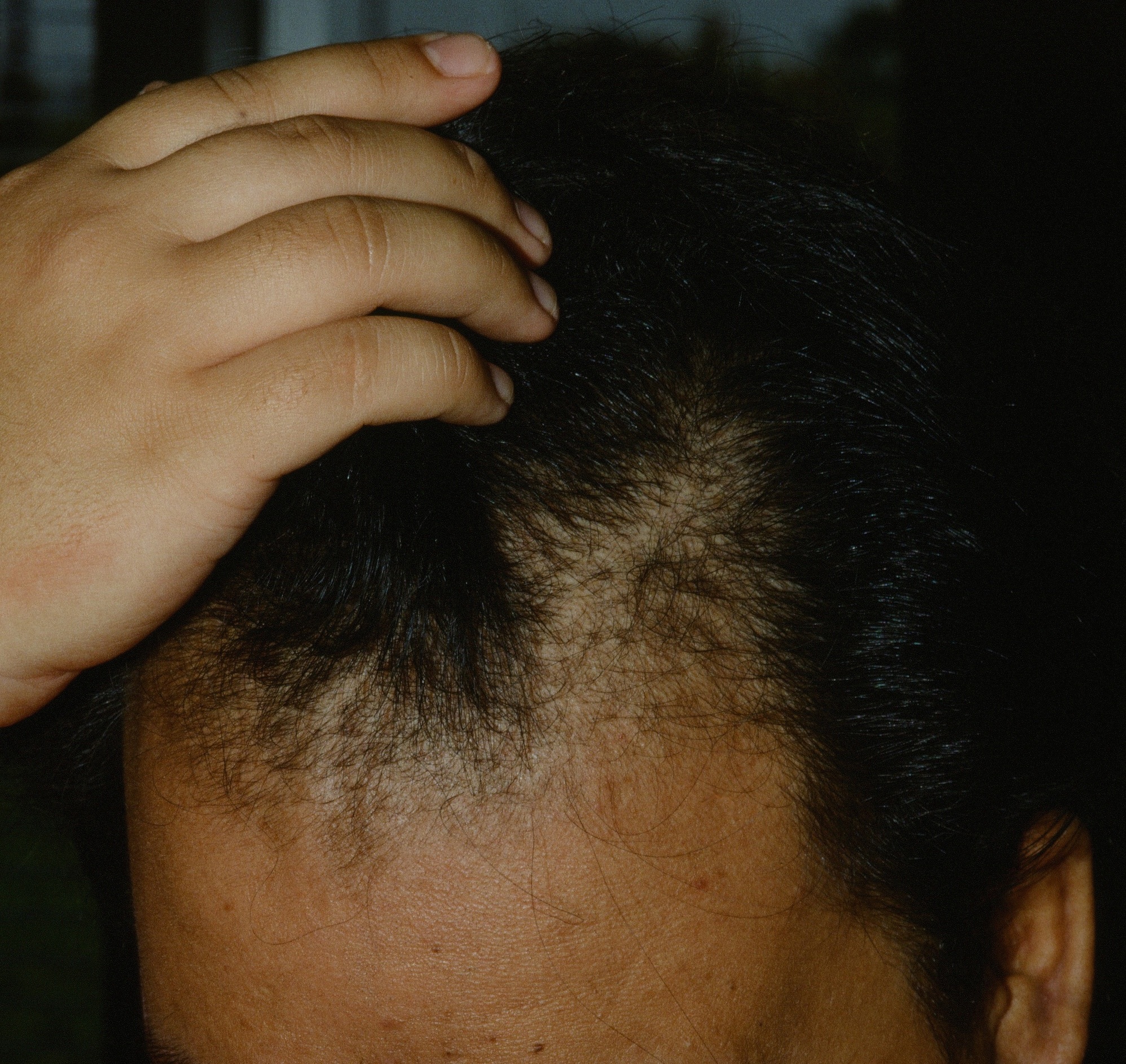
All labs normal. Female pattern hair loss.
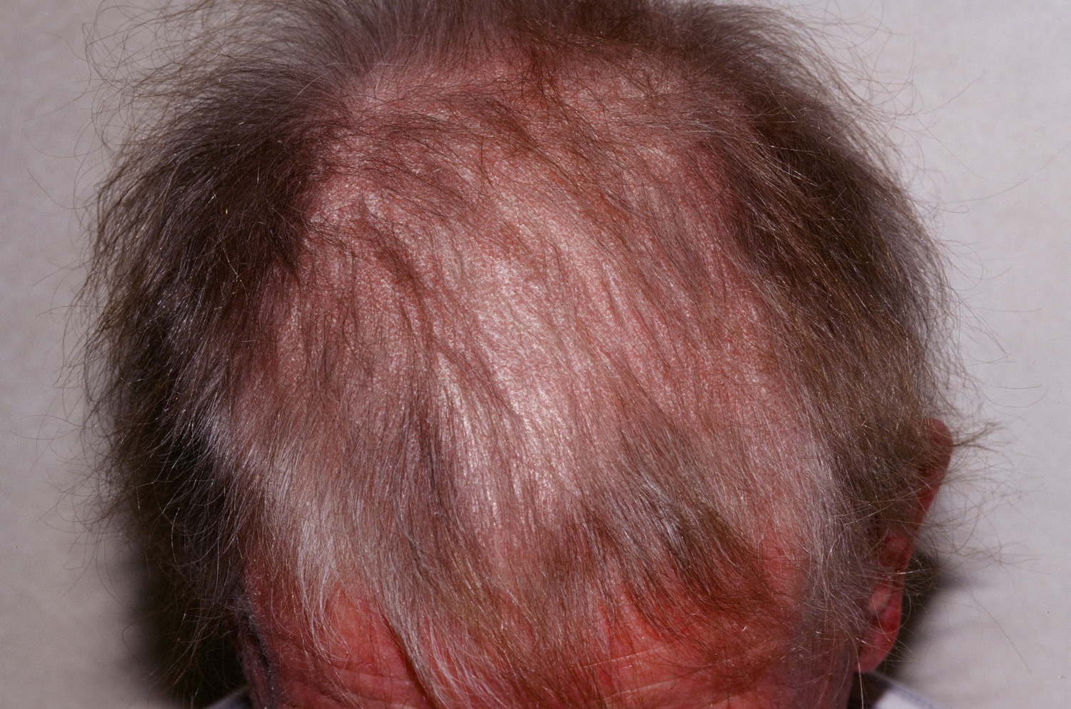
This woman with systemic lupus had significant hair loss. Her disease was obvious as is usually the case.
Homepage | Who is Dr. White? | Privacy Policy | FAQs | Use of Images | Contact Dr. White