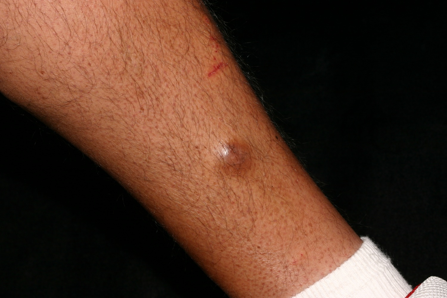
 A flesh-colored to reddish/brown bump on the leg is typical.
A flesh-colored to reddish/brown bump on the leg is typical.
The dermatofibroma is a very common benign fibrous growth or tumor of the skin, occurring most commonly on the leg.
These dermal papules most commonly present on the leg of an adult woman. They are usually brown, but may be flesh-colored or black. A variant called a Hemosiderotic Dermatofibroma may contain significant iron and appear blue/black. Sometimes they occur on the upper back or arm. Squeezing them between the thumb and forefinger produces a dimpling that is characteristic. Multiple dermatofibromas may rarely occur.
The atrophic dermatofibroma is characterized by a flat or atrophic and depressible surface. It is frequently seen on the back of a woman. Histopathological features show reduction of the thickness of the dermis and elastic fibers. It has been suggested that the atrophic DF represents the end-stage DF. This concept is reinforced by the association of older age with atrophic DF.
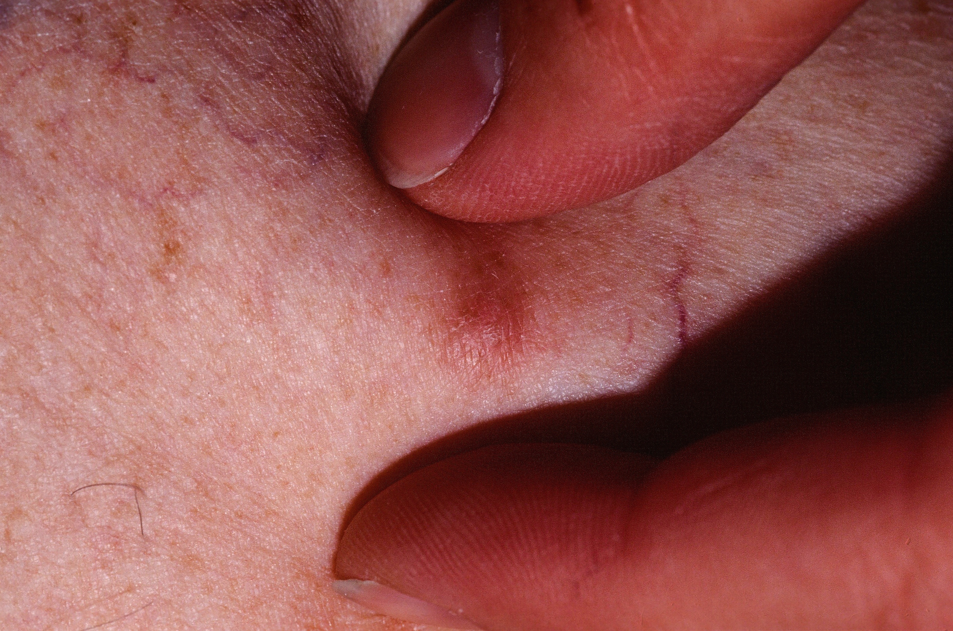
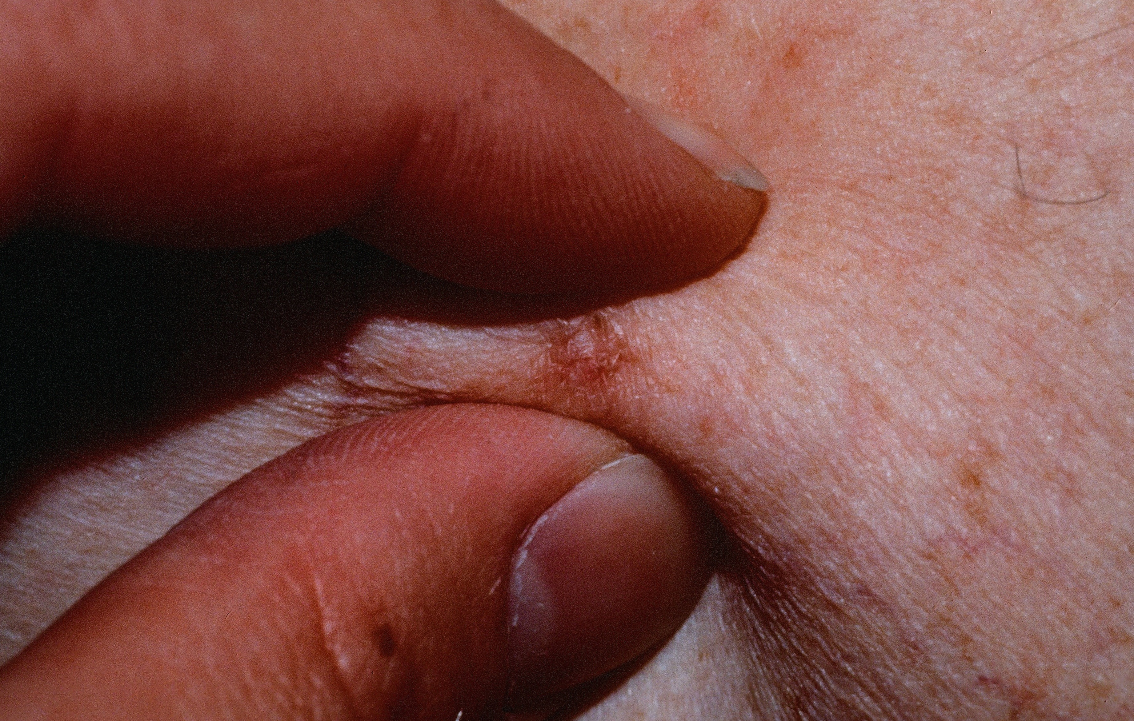
To see if a brown lesion is a dermatofibroma (DF), squeeze it. If you can feel a dermal thickening, and the surface pulls in, it is.
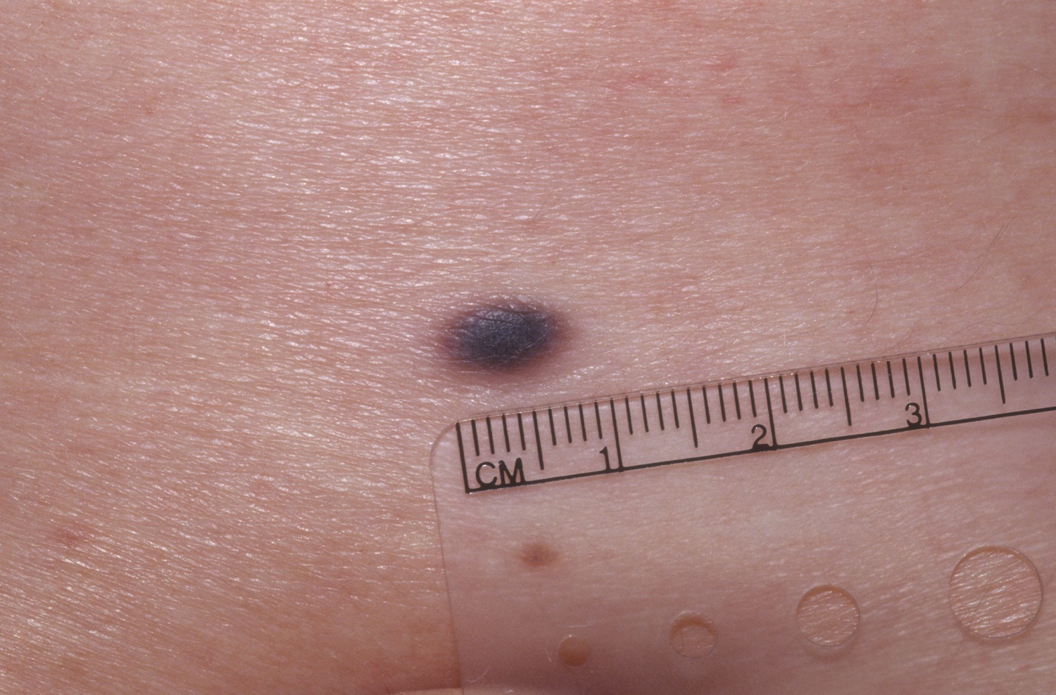
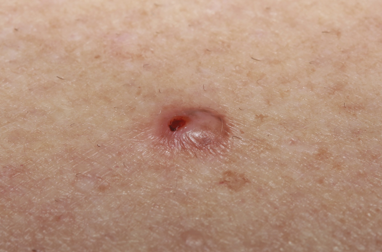
The DF may be darkly pigmented.
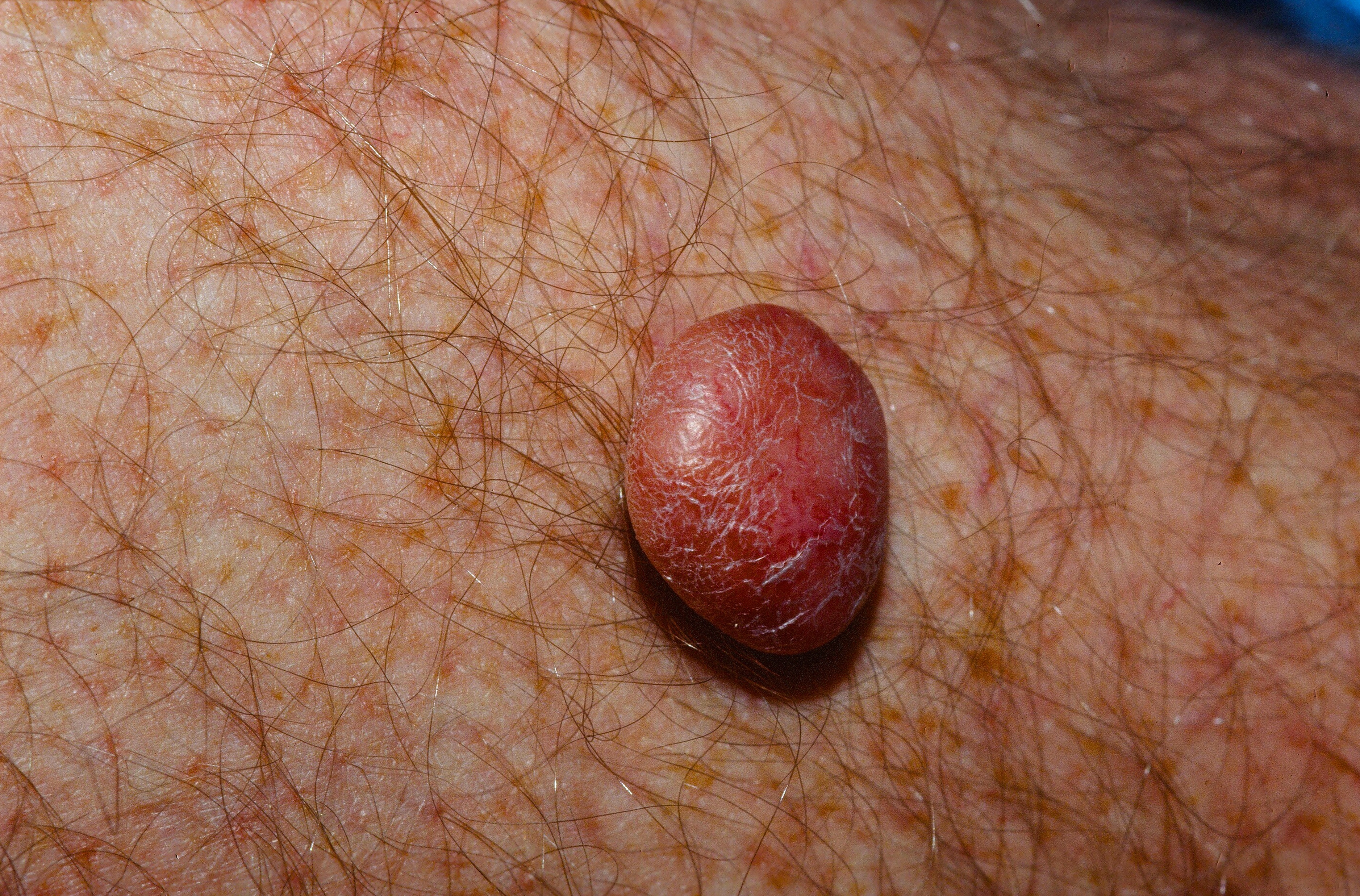
Pedunculated or papulonodular dermatofibromas.
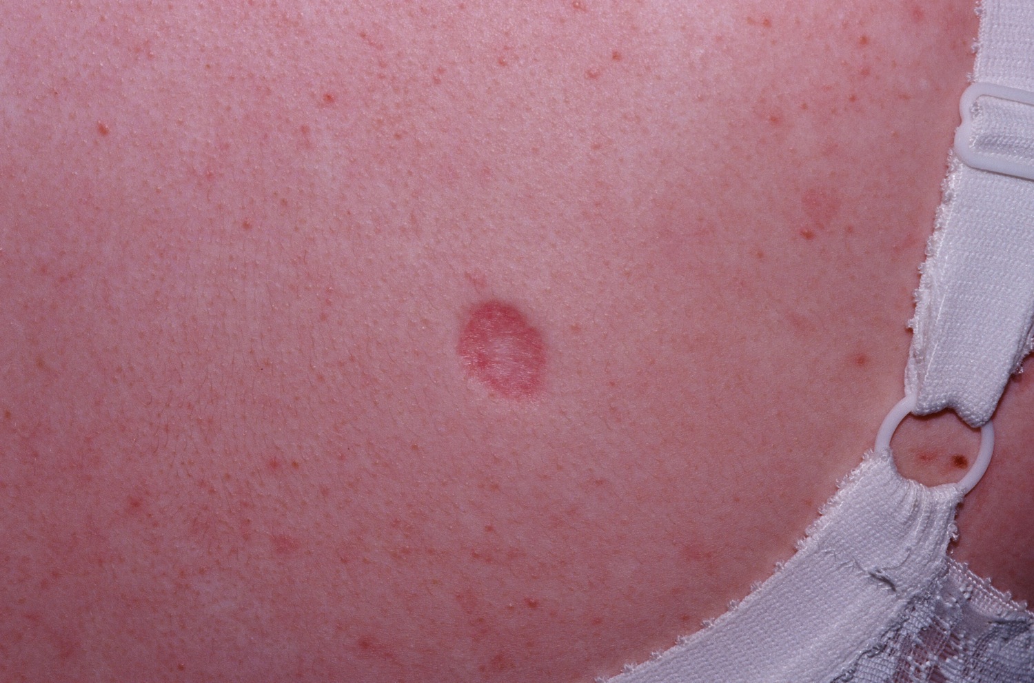
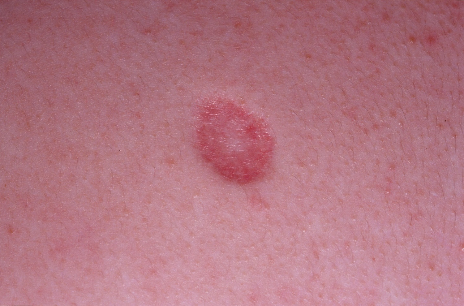
The atrophic variant of the dermatofibroma, common on the back.
This raised DF on a woman's leg was constantly cut shaving. A scoop shave was performed with excellent results.
The term fibrous histiocytoma is sometimes used for larger, nodular lesions analogous to the dermatofibroma.
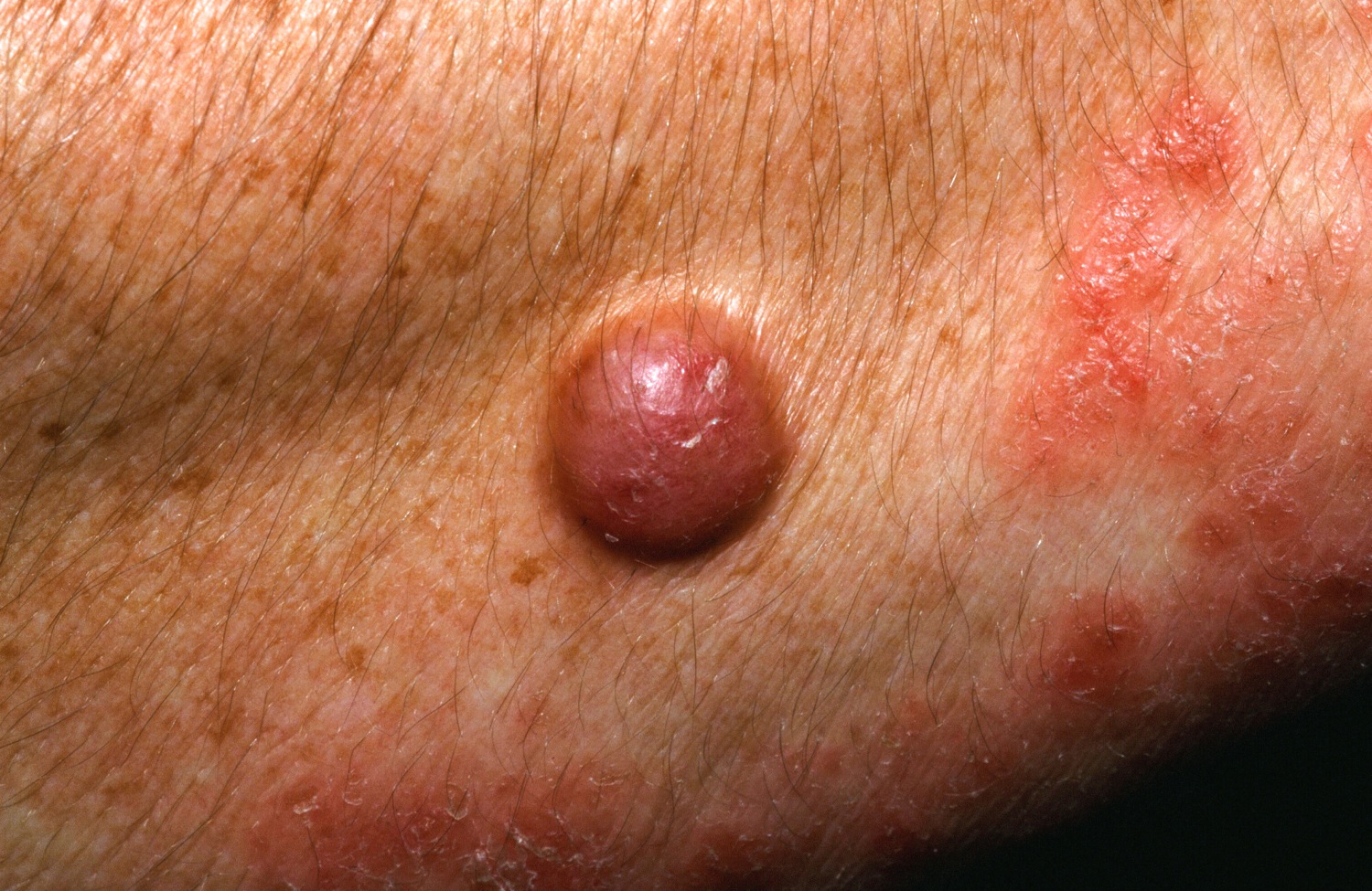
A DF on the arm in a patient with psoriasis.
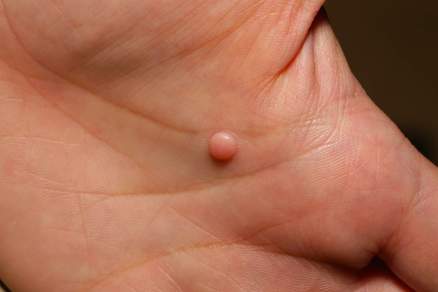
A DF in an unusual location (the palm).
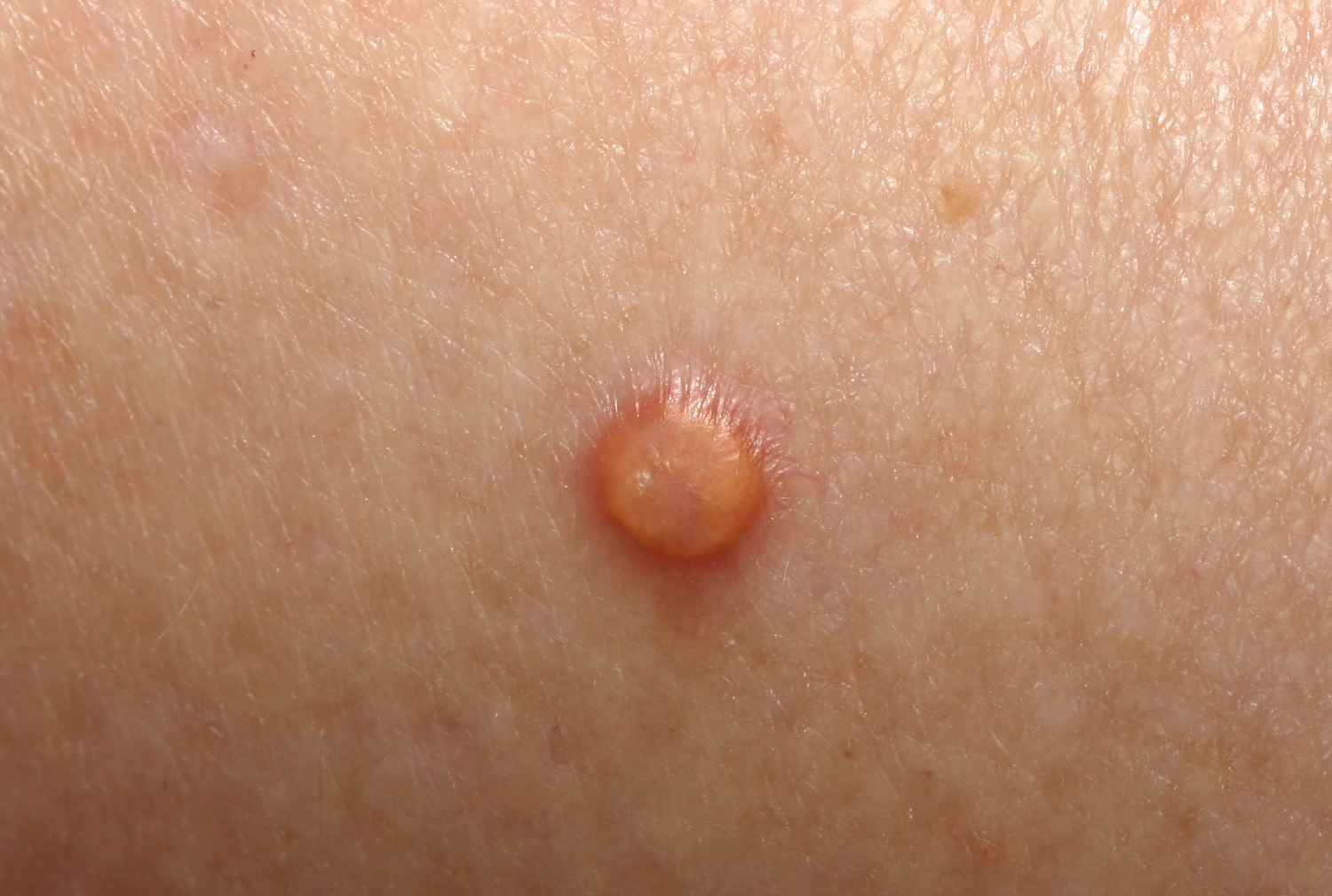
An unusually yellow DF (biopsy-proven).
Homepage | Who is Dr. White? | Privacy Policy | FAQs | Use of Images | Contact Dr. White