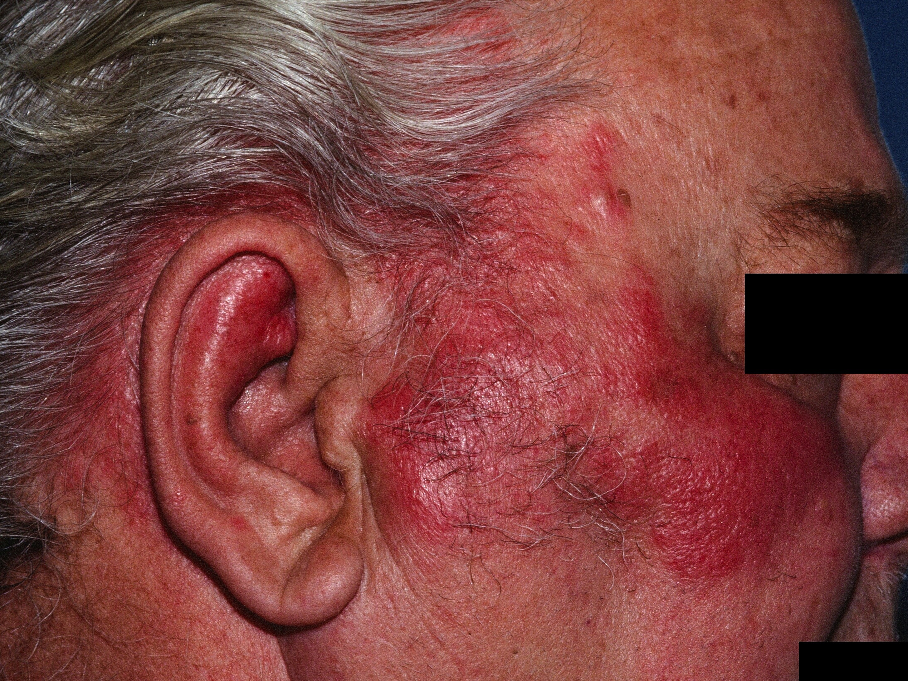
This man developed a fever and this rash over 2 days. He was admitted for IV antibiotics. Cellulitis of the midface is particularly dangerous because of its proximity to vital structures, including the eyes and brain.
This man developed a fever and this rash over 2 days. He was admitted for IV antibiotics. Cellulitis of the midface is particularly dangerous because of its proximity to vital structures, including the eyes and brain.
Cellulitis is a rapidly expanding bacterial infection of the skin. Impetigo is also a bacterial infection of the skin but is primarily confined to the superficial layers. Cellulitis involves deeper layers of the skin including the dermis. Erysipelas is a specific subset of cellulitis caused by streptococcus. See also bacterial infections of the skin here.
Clinically, one sees a warm, edematous, expanding plaque. The face and lower legs are common sites. On the lower legs, confusion with stasis dermatitis often occurs.
Necrotizing fasciitis is a more severe infection that can be life-threatening. It may be suspected when there are systemic signs (e.g., fever, chills) or if the skin shows more than just erythema and edema, e.g., hemorrhage, necrosis, or fluctuance.
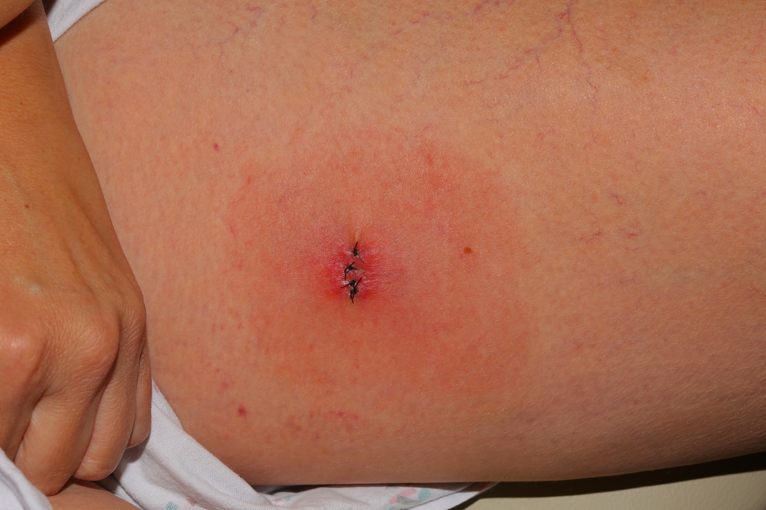
This patient called 4 days after minor surgery to say she thought her wound was infected. It was!
Homepage | Who is Dr. White? | Privacy Policy | FAQs | Use of Images | Contact Dr. White