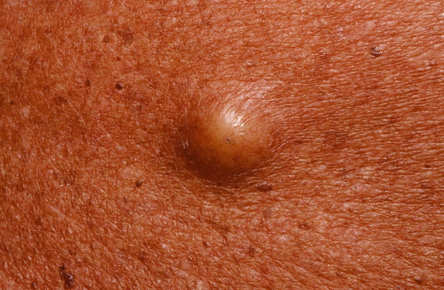
A dermal nodule 0.5-2.0 cm in diameter is typical. A central pore is usually visible. The patient often relates being able to squeeze out a foul-smelling material.

A dermal nodule 0.5-2.0 cm in diameter is typical. A central pore is usually visible. The patient often relates being able to squeeze out a foul-smelling material.
The epidermal inclusion cyst (EIC)--formerly known as the sebaceous cyst--is a sphere of skin within the skin. The cyst wall constantly flakes into the center of the cyst causing it to enlarge over time.
A dermal nodule 0.5-2.0 cm in diameter is typical. A central pore is usually visible. The patient often relates being able to squeeze out a foul-smelling material. If left alone, the lesion may at some point become acutely inflamed. This is usually is a result of rupture of the cyst wall and release of the cyst contents into the surrounding dermis. A foreign body response results. The patient then presents with an acutely inflamed and painful nodule where the asymptomatic bump used to be. Initially, the lesion is firm, but later it becomes fluctuant and may ultimately drain. The skin heals having extruded the cyst. Alternatively, the inflammation may subside and the lesion returns to an asymptomatic bump. When patients present this way, the clinician often assumes the lesion is "infected". Cultures usually show it is not.
In one review of 13,746 EICs, 48 contained a malignancy, for an incidence of 0.3% and with the most common malignancy being squamous cell carcinoma
An EIC of the ear.
Who is Dr. White? | Privacy Policy | FAQs | Use of Images | Contact Dr. White